Indwelling urinary catheters are still one of the most commonly used clinically invasive devices across the UK (Loveday et al 2014; Feneley et al, 2015). It has been estimated that at least 90,000 people living in community settings across England currently use urinary catheters long term (Gage et al, 2017), and almost one-quarter of all inpatients experience an indwelling catheter at some point during their hospital stay (Health Protection Scotland, 2012; Feneley et al, 2015).

Evidence over several years has indicated that the length of time a catheter is in place has a direct implication on the increased risk of developing an infection, with figures of 20% widely referenced (Chenoweth and Saint, 2013; Loveday et al, 2014). It is recognised that a significant number of these individuals will develop sepsis, and in some cases lead to fatalities — estimated to be around 2100 deaths a year directly caused by the use of indwelling catheters (Feneley et al, 2015). Indwelling catheters are potentially risky devices and have an adverse effect on the health and social wellbeing of individuals, as well as a financial implication for services.
 

‘Indwelling catheters are potentially risky devices and have an adverse effect on the health and social wellbeing of individuals, as well as a financial implication for services.’


The current cost of treating catheter-associated urinary tract infections (CaUTIs) is reported to be almost 2000 an episode (Yates, 2016), and the total cost of using Foley catheters within the NHS is estimate to be between 1.0 and 2.5 billion a year (Yates, 2016).

While it is recognised that an indwelling catheter is a last resort, there are several valid reasons for their use (Geng et al, 2012; Royal College of Nursing [RCN], 2019; Table 1). Any situation where the bladder cannot effectively empty or drain should be carefully assessed and consideration taken as to how to manage the situation and relieve symptoms safely and effectively. Use of indwelling catheters should be planned around an individual’s needs and appropriate management and early removal must be addressed. Questions are often raised regarding the optimum time to perform a trial without catheter (TWOC). This article will explore the optimum timing for TWOC and current evidence advising on how to safely and effectively manage a TWOC.

In health care we are guided by best practice and clinical evidence. The RCN’s best practice guidelines relating to catheterisation were recently revised (RCN, 2019). They are aimed at any healthcare professional responsible for safe and effective catheterisation techniques and should be readily available and widely evidenced in everyday practice across the various health and social care environments. NHS Improvement also released national clinical documentation to support the safe and effective use of urinary catheters (NHS Improvement, 2019). The most recent evidence-based guidelines reflect changes in practice and healthcare culture, and demonstrate that registered and non-registered staff are potentially responsible for insertion and removal of catheters (RCN, 2019). Any healthcare professional participating in elements of catheter care, including insertion and removal, should be able to demonstrate safe effective practice in line with current clinical evidence.

PURPOSE OF TWOC AND WHEN IT SHOULD BE CONSIDERED?

The primary aim of a TWOC is to establish the ability to effectively and successfully empty the bladder unaided (Robinson, 2005; Table 2). A TWOC should always be planned in advance with consideration around:
  • Timing of catheter removal
  • The environment in which the TWOC is performed
  • Ongoing assessment of bladder function, including any retention
  • A clear plan of action should the TWOC be considered a failure (Yarde, 2015; Bardsley 2017).
There is no substantive clinical evidence on the best time to plan a TWOC, but anecdotally two weeks post catheter insertion (for urine retention) has been a popular choice. One clinical paper suggested a TWOC should ideally be performed within two to three days after initial catheterisation when the reason for the catheter has been acute urine retention (Kuppusamy and Gillatt, 2011). The paper reports a 40% success rate for TWOCs in patients whose underlying cause of acute urinary retention (including constipation, urinary tract infection [UTI] and medication effects) has been resolved.

TWOCs may also be more successful in early stages for those patients with a residual urine volume of less than one litre and no previous lower urinary tract symptoms (LUTs) (Emberton and Fitzpatrick, 2008). Planning a TWOC when the catheter has been inserted for other reasons has also been the subject of much discussion and practice variation, with some areas taking catheters out in the early morning (Addison, 2001; Warering, 2001; Warrilow et al, 2004) and others preferring midnight (Kelleher, 2001). There is no substantive clinical evidence to support removing a catheter at a particular time of day in relation to a more or less successful TWOC.

The environment in which a TWOC is carried out may also have a bearing on success. In the author’s clinical experience, TWOC clinics in healthcare environments can provide close supervision and formal management of removal of a catheter and achieve optimum outcomes for patients with support from healthcare professionals. A successful TWOC may be determined over several hours, so attendance at an acute unit can become timely and costly as a patient would need to remain on the unit until the TWOC outcome has been determined. However, it is now commonplace to offer a TWOC within a patient’s own home, as long as a suitably experienced healthcare professional is available to monitor and support the patient to determine the success of the procedure, with a clear follow-up protocol in place (Gilbert, 2006).

The author has not found any substantive clinical evidence to suggest a particular time of day, or time from insertion of catheter to removal, improves the outcome or success of a TWOC. The author’s recommendation is that the timing of a TWOC should be based on individual assessment of need, environment and follow-up plans to ensure effective independent bladder emptying is re-established. The reason for initial catheterisation and planned strategies to manage a possible failed TWOC are also important elements to consider.

table1


Table 2

WHO MAKES THE DECISION TO TWOC?

The presence of an indwelling urinary catheter is associated with an increased risk of infection; this has been widely evidenced and discussed. The use of evidence-based nurse-led daily checklists can decrease CaUTI rate significantly (Fuchs et al, 2011; Yatim et al, 2016; Purvis et al, 2014). The HOUDINI protocol was developed by Adams et al (2012) and is a nurse driven protocol offering staff a method of determining when to remove a catheter. The acronym stands for:
  • Haematuria, gross
  • Obstruction (urinary)
  • Urology surgery
  • Decubitus ulcer — category 3 or 4, or open sacral or perineal wound in incontinent patient
  • Input and output measurement for patient management or hemodynamic instability
  • Nursing — end-of-life care
  • Immobility due to physical constraints.
The protocol recommends a daily challenge of asking whether the patient meets any of the criteria reflected in the acronym. When a patient no longer meets the criteria, the catheter should be removed. The HOUDINI protocol provides a structure enabling nurses to make decisions and ensure safe and quality patient care. Introducing a protocol into an organisation can have challenges and needs careful planning and structure that offer training and support to staff to effectively implement the tool. The protocol is widely included in many organisational policies on catheterisation and has been referenced in the national RCN (2019) guidelines on best practice.

DOES THE DRAINAGE EQUIPMENT USED HAVE AN IMPACT ON THE SUCCESS OF TWOC?

Normal bladder filling and storage involve the bladder stretching to accommodate a reasonable volume of urine (it is generally accepted that a normal bladder capacity in an adult is 400–600mls). The detrusor muscle is at rest until the bladder capacity for the individual is reached, whereby a series of nerve impulses communicate between the bladder and brain to start the emptying phase. During the filling and storage phase, the urethra and sphincters must remain closed and the pelvic floor contracted to ensure no leakage occurs. During the emptying phase, the detrusor contracts, the urethra and sphincters open and the pelvic floor relaxes, and the intravesical pressure increases to effectively empty the bladder contents.

Bladder function in some patients can slow down or alter over the time a catheter is in situ, especially if the urine is on continuous drainage into a bag (Addison, 2001; Robinson, 2005). Use of continuous drainage increases the negative pressure inside the bladder, creating a suction-like effect that may result in trauma of the submucosal bladder lining and urethra or supra pubic tract (Feneley et al, 2015). The bladder tone may also be affected with a free drainage catheter — the recognition of the normal bladder filling and emptying cycle is interrupted as the free drainage system does not allow urine to expand the bladder.

Use of a catheter valve system mimics the filling and emptying phases and therefore may be an indicator for a more successful TWOC (Woodward, 2013; Yates, 2016). It is thought that the valve system helps to preserve some sensation and detrusor function by mirroring a more normal micturition pattern.

Commonly in practice, the timescale to wear a valve system appears to range from 48 hours to two weeks before removing the catheter, although there is no substantive clinical evidence to support this. However, allowing the bladder to maintain a filling and emptying cycle through the use of valve devices will in all probability improve the outcome of TWOC. The clinician should follow local policy and guidelines for use of catheter valves, as it should be noted some patients cannot safely use a catheter valve (see Table 3 for indications and contraindications for catheter valve use). These patients can still, however, have a successful TWOC procedure with planning and monitoring.


POTENTIAL ISSUES TO CONSIDER WITH TWOCs

Removing an indwelling urinary catheter is a relatively simple and uncomplicated procedure. However, there are some situations that may present a challenge for clinicians, such as:
  • Some patients will experience an increased sense of pain or discomfort when the catheter is removed. This may be caused by the negative pressure of an empty bladder, as previously stated. Consideration of timing of the TWOC procedure, maintaining a partially filled bladder immediately before removing the catheter may reduce discomfort
  • The clinician may not be able to fully deflate the retention balloon of the catheter. This may be due to a faulty or damaged valve on the catheter, stone formation or encrustation, or debris in the bladder. Careful assessment, including the clarity of the urine, any issues identified with drainage during the lifetime of the catheter, dehydration and bowel activity may offer potential solutions to prevent these issues from occurring again
  • Latex and silicone are commonly used in catheter manufacture. The catheter balloon often has residual cuffing, ridging or wrinkling as the balloon is deflated, due to the nature of expansion and contraction of the materials over the timescale the catheter is worn. This creates uneven rough edges causing the catheter to stick to the urethral or suprapubic tract on removal, thereby increasing the risk of trauma and infection. The catheter balloon volume also has often decreased over the time a catheter is in situ through natural osmosis. Fully deflating the balloon and then re-inflating with 1ml of water will help to smooth out the rough edges and enable easier removal — use of extra lubricant will also reduce the trauma risk on removal. It should be noted that under normal circumstances catheter balloons should only be inflated once on insertion and deflated once on removal.
The European Association of Urology Nurses (EAUN) provided a comprehensive troubleshooting guide within their best practice guidelines, with management strategies for dealing with issues during the TWOC (Table 4) (Geng et al, 2012).
Table 3
Table 4

ESSENTIAL MONITORING WITH TWOC PROCEDURE

An agreed time should be established for removal of the catheter. This should take into account any follow-up required to establish a successful outcome. This may vary dependent on whether the patient is at home or in an environment where there are healthcare professionals available during the whole process. Once a catheter has been removed, the patient’s urine output and fluid intake should be monitored over a period of time to ensure that the bladder emptying pattern is consistent and effective. This may be the responsibility of the patient or a family member. Therefore, the person removing the catheter will need to specify exactly what information should be recorded and how. Ideally, a healthcare professional should reassess the patient at an appropriate time interval and carry out bladder scanning to check residual volumes (Burkhard et al, 2016). This is to confirm whether normal emptying and filling has been re-established.

As normal bladder filling and emptying takes place for most people over a 6–8-hour period, healthcare professionals should schedule a scan within this timescale. In some cases, it may be acceptable to wait up to 12 hours before scanning, but this will be based on individual assessment of clinical symptoms. It should be noted that to achieve accurate post void residual (PVR) measured on a bladder scanner, the scan must take place within twenty minutes of the spontaneous void (Newman, 2007).

The general opinion of urologists worldwide when considering urine retention is a PVR of <100mL is not significant, while if the PVR is >100mL, it should be investigated further to establish a pattern of retention (Gallien et al, 2005; Ghalayini et al, 2005). It is important to understand the significance of the residual volume, the spontaneous void volume, and the fluid intake of the patient to be able to accurately interpret the results and make a decision as to whether the TWOC has been a success, or if the patient needs re-catheterising.

WHAT SHOULD A PATIENT EXPECT POST TWOC?

Urgency and frequency of micturition following a TWOC are also reported by some patients and may be caused by a residual inflammation of the urethra from the presence of a catheter or by an infection. Patients with bothersome symptoms should be tested for a UTI and treated according to clinical presentation and local policy. Overactivity of the detrusor should be eliminated as a cause of the frequency and urgency, as this can be successfully treated in some patients with anticholinergic medication (Hanno et al, 2011; Feneley et al, 2015).

Bladder capacity may have been reduced following long-term use of a catheter, particularly if the catheter has been on free drainage. This will require a structured programme of bladder retraining once a TWOC has been performed. The retraining programme needs to be individualised and take into account patient dietary and fluid intake, as there are some well-known irritants such as caffeine, nicotine, carbonated drinks and artificial sweeteners that can adversely affect the bladder (Colley, 2015). Timed voiding is a recognised method of retraining the bladder, based on the amount a normal healthy bladder should hold (400–600mls in adults), and how many times a day the normal healthy bladder empties (4–8 times in 24 hours). This can establish a ‘normal’ bladder function even in the absence of sensation, with voiding set to times and prompted throughout the day. Distraction techniques when the sensation to void is frequent, building up the intervals between voiding over a period of days can increase bladder capacity and help regain a more normal pattern of micturition (Colley, 2015).

Incomplete emptying of the bladder should be determined through the use of frequency volume charts (Colley, 2015; Holroyd, 2020), and bladder scans post voiding at appropriate intervals post catheter removal. Clinicians should refer to local policy on how and when to perform a post void bladder scan following a TWOC procedure.

It is recognised that patients who experience complications in relation to TWOC and catheters would benefit from specialist nursing input to ensure that the best management plan and options are considered and implemented (Marvin and Mills, 2015).

A failed TWOC is determined when a patient fails to void successfully, effectively resulting in a need to re-catheterise (Gilbert, 2006). If a patient repeatedly fails a TWOC procedure, or has repeated episodes of urinary retention, urology opinion should be sought. Clinicians should refer to local policy for guidance on the referral pathway in their locality.

‘Clinical evidence concludes that where possible the catheter should be removed at the earliest opportunity to reduce the risk of CaUTI.’


CONCLUSION

Indwelling urinary catheters are a high risk intervention, but unavoidable in some situations. Indwelling catheterisation should only be considered when all other options have been discounted (Geng et al, 2012; RCN, 2019; NICE, 2014). There should be evidence of comprehensive and individualised patient assessment with a clear documented reason for the catheter and a review or removal date (Simpson, 2017; Geng et al, 2012; RCN, 2019; Loveday et al, 2014). The impact of catheter complications is felt by the patient who suffers the often significant issues relating to infection and antibiotic use (Chapple et al, 2016).

Nursing and care services, both in community and emergency departments, experience increased demand on their services, a reduction in capacity and ability to respond to the holistic needs of patients with indwelling catheters, resulting in a rapidly increasing economic burden on the NHS and social care sector (Tay et al, 2016; Ansell and Harari, 2017).

Clinical evidence concludes that where possible the catheter should be removed at the earliest opportunity to reduce the risk of CaUTI. Daily reviews of any indwelling catheter and the need for them are recommended (Andreessen et al, 2012), with a structured system or protocol to manage the TWOC (Carter et al, 2014; Meddings et al, 2014; Purvis et al, 2014). Planning and implementing a TWOC procedure should consider many factors that could affect the success of the TWOC.
This piece was first published in the Journal of Community Nursing. To cite this article use: Holroyd S (2020) Trial without catheter: what is best practice? J Community Nurs 34(2): 60-67

References

Adams D, Bucior H, Day G, Rimmer JA. (2012) HOUDINI: make that urinary catheter disappear — nurse-led protocol. J Infect Prevent 13(20): 44–6

Addison R (2001) Practical procedures: trial removal of a catheter. Nurs Times 97(4): 14–15

Andreessen L, Wilde MH, Herendeen P (2012) Preventing catheter-associated urinary tract infections in acute care: the bundle approach. J Nurs Care Qual 27(3): 209–17

Ansell T, Harari D (2017) Urinary catheter-related visits to the emergency department and implications for community services. Br J Nurs 26(9 suppl): S4–11

Bardsley A (2017) How to remove an indwelling urinary catheter in female patients. Nurs Standard 31(19): 42–5

Burkhard FC, Lucas MG, Berghmans LC, Bosch JLHR, Cruz F, Lemack GE, Nambiar AK, Nilsson CG, Pickard R, Tubaro A (2016) EAUN Guidelines on Urinary Incontinence in Adults. European Association of Urology Nurses, the Netherlands

Carter NM, Reitmeier L, Goodloe LR (2014) An evidence based approach to the prevention of catheter-associated urinary tract infections. Urologic Nurs 34(5): 238–41

Chapple A, Prinijha S, Feneley R, Ziebland S (2016) Drawing on accounts of longterm urinary catheter use: design for the seemingly mundane. Qual Health Res 26(2): 154–63

Chenoweth C, Saint S (2013) Preventing catheter-associated urinary tract infections in the intensive care unit. Crit Care Clin 29(1): 19–32

Colley W (2015) Use of frequency volume charts and voiding diaries. Nurs Times 11(5): 12–15

Emberton M, Fitzpatrick J (2008) The Reten- World survey of the management of acute urinary retention: preliminary results. BJU Int 101(Suppl 3): 27–32

Feneley R, Hopley I, Wells P (2015) Urinary catheters history: current status, adverse events and research agenda. J Med Eng Technol 39(8): 459–70

Fuchs MA, Sexton DJ, Thornlow DK, Champagne MT (2011) Evaluation of an evidence-based, nurse-driven checklist to prevent hospital-acquired catheterassociated urinary tract infections in intensive care units. J Nursing Care Qual 26(2): 101–9

Gage H, AveryM, Flannery C, Williams P, Fader M (2017) Community prevalence of long-term urinary catheters use in England. Neurourol Urodyn 36(2): 293–6

Gallien P, Reymann J, Amarenco G, Nicolas B, de Se`ze M, Bellissant E (2005) Placebo controlled randomized double blind study of the effects of botulinum A toxin in detrusor sphincter dyssynergia in multiple sclerosis patients. J Neurol Neurosurg Psychiatry 76: 1670–3

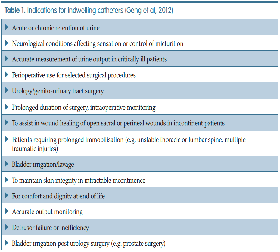
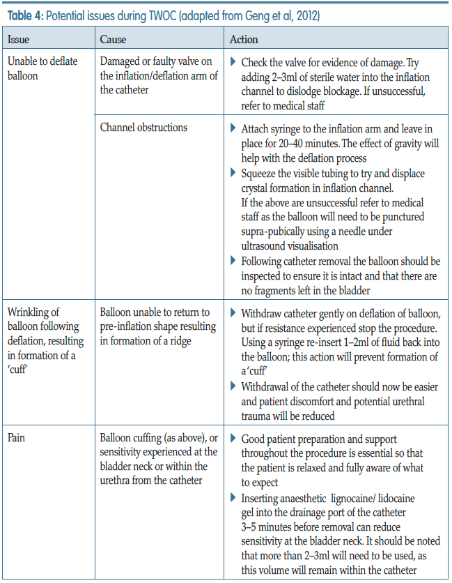
Geng V, Cobussen-Boekhorst H, Farrell J (2012) Catheterisation: indwelling in adults. European Association Of Urology Nurses, The Netherlands

Ghalayini I, Al-Ghazo M, Pickard R (2005) A prospective randomized trial comparing transurethral prostatic resection and clean intermittent self-catheterization in men with chronic urinary retention. BJU Int 96: 93–7

Gilbert R (2006) Procedure to undertake a trial without catheter. Nurs Times 102(42): 48–50

Hanno PM, Burks DA, Clemens JQ (2011) AUA guideline for the diagnosis and treatment of interstitial cystitis/bladder pain syndrome. J Urol 185(6): 2162–170

Health Protection Scotland (2012) Scottish National Point Prevalence Survey of Healthcare Associated Infection and Antimicrobial Prescribing 2011. Available online: http://bit.ly/1SJCIcK

Holroyd S (2020) Frequency volume charts and fluid balance monitoring: getting it right. J Community Nurs 34(1): 40–3

Kelleher M (2001) Removal of urinary catheters: midnight vs 06.00 hours. Br J Nurs 11(2): 84–90

Kuppusamy S, Gillatt D (2011) Managing patients with acute urinary retention. Practitioner 255(1739): 21–3, 2–3

Loveday HP, Wilson JA, Pratt RJ (2014) EPIC 3: national evidence-based guidelines for preventing healthcare-associated infections in NHS hospitals in England. J Hosp Infect 86(suppl 1): S1–S70

Feneley R, Hopley I, Wells P (2015) Urinary catheter history: current status, adverse events and research agenda. J Med Eng Technol 39(8): 459–70

Mavin C, Mills G (2015) Using quality improvement methods to prevent catheter-associated UTI. Br J Nurs Urology Supplement 24(18): S22–S28

Meddings J, Rogers MA, Krein SL, Fakih MG, Olmsted RN, Saint S (2014) Reducing unnecessary urinary catheter use and other strategies to prevent catheter-associated urinary tract infection: an integrative review. BMJ Qual Saf 23: 277–89

National Institute for Health and Care Excellence (2014) Long-term urinary catheters: prevention and control of healthcare associated infections in primary and community care. NICE, London

NHS Improvement (2017) Preventing healthcare associated Gram-negative bloodstream infections: an improvement resource. Public Health England, London

NHS Improvement (2019) Urinary catheter tools. Available online: https:// improvement.nhs.uk/resources/urinarycatheter- tools/

Newman DK (2007) The indwelling urinary catheter: principles for best practice. J Wound Ostomy Continence Nurs 34(6): 655–61

Purvis S, Gion T, Kennedy G, Rees S, Safdar N, VanDenBergh S, Weber J (2014) Catheter-associated urinary tract infection: a successful prevention effort employing a multipronged initiative at an academic medical center. J Nurs Care Qual 29(3): 141–8

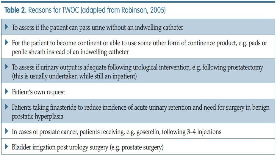
Robinson J (2005) Removing indwelling catheters: trial without catheter in the community. Br J Community Nurs 10(12): 553–4, 556–7

Royal College of Nursing (2019) Catheter Care: RCN Guidance for nurses. RCN London

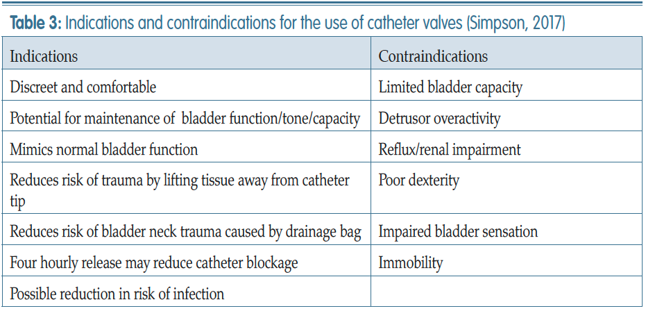
Simpson P (2017) Long-term urethral catheterisation: guidelines for community nurses. Br J Nurs 26(9 suppl): S22–26

Tay I J, Lyons H, Karrouze I, Taylor C, Khan AH, Thompson PM (2016) Impact of the lack of community catheter care services on the emergency department. BJU Int 118(2): 32734

Warering M (2001) Proving a domiciliary urology service. Professional Nurse 12(3): 169–75

Warrilow M, Williams D, Guest J (2004) The introduction of a trial without catheter model in primary care. Br J Nurs 13(7): 1035–40

Woodward S (2013) Catheter valves: a welcome alternative to leg bags. Br J Nurs 22(11): 650, 652–4

Yarde D (2015) Managing indwelling urinary catheters in adults. Nurs Times 111(22): 12–13

Yates A (2016) Indwelling urinary catheterisation: what is best practice? Br J Nurs 25(9) urol suppl: S4–12

Yatim J, Wong K, Ling M, Tan S, Tan K, Hockenberry M (2016) A nurse-driven process for timely removal of urinary catheters. Int J Urol Nurs 10(3): 167– 72