Moisture-associated skin damage (MASD) is an umbrella term to describe the trauma that occurs when there is prolonged exposure to moisture. Incontinence-associated dermatitis (IAD) is an MASD condition caused by urinary and/or faecal incontinence. It often causes significant pain and has a negative impact on an individual’s quality of life. Prompt diagnosis of IAD is key to successful management, but the early symptoms of erythema and redness may not be obvious in those with a dark skin tone. Delays in assessment and treatment mean that maceration, infection and the development of pressure ulcers become more of a risk. This is compounded by the lack of education and awareness of the significance of skin tone in relation to wound care. This article is based on published research, together with the practical experience of healthcare professionals at the Complex Wound Clinic (CWC) in North West London. The aim is to highlight the importance of considering skin tone when managing IAD. This can be achieved by using a validated skin tone assessment tool which shows a range of six skin tones. The importance of skin tone is a critical subject as the latest 2021 Census data shows that in England and Wales, 18% belong to a black, Asian, mixed or other ethnic group (Gov.UK, 2021 Census data).  

THE COMPLEX WOUND CLINIC  


The Complex Wound Clinic (CWC) at Central and North West London (CNWL) NHS Foundation Trust provides wound assessment and management to help improve chronic wound healing rates.  

The specialist team assist other clinicians in the care of patients with complex wounds, providing the expert support required to deliver the most appropriate management. The service is provided in the community, in patients’ homes (including nursing homes) and in clinics. Since the Covid pandemic, an increasing number of patients are being referred with moisture-associated skin damage (MASD), mostly due to incontinence-associated dermatitis (IAD). Many patients have had little or no previous contact with healthcare services; preferring to keep their continence problems ‘in the family’.  

THE SKIN  


The skin is the largest organ of the body, providing a protective barrier against pathogens. It also protects internal tissues and organs from harmful ultraviolet (UV) radiation, chemical irritants, temperature, toxins, and mechanical injuries (Lopez-Ojeda et al, 2022). To maintain this protective function, skin integrity is essential. When the skin is exposed to excess moisture, it becomes prone to maceration and breakdown (Green et al, 2022). This may lead to the development of complex problems, including pressure ulceration, MASD, skin tears and infections (Waller and Cole, 2023).  

MOISTURE-ASSOCIATED SKIN DAMAGE (MASD)  


MASD is caused by prolonged exposure of the skin to moisture. This compromises the protective function of the skin, causing it to become more susceptible to penetration by microorganisms, as well as mechanical damage from shear and friction (Gray et al, 2011).  

MASD can be aggravated by chemical irritants, proteolytic and lipolytic enzymes and an alteration in the skin pH (Young, 2017), all of which have the capacity to compromise the barrier function. Damage can range from superficial erythema to extensive skin breakdown and may be complicated by bacterial and fungal infections (Young, 2017). It is more frequently seen in older skin, which is more fragile and susceptible to breakdown. With increasing age, epidermal turnover becomes slower, there are fewer sweat glands and sebum production is reduced (Young, 2017).  

Additional risk factors for skin breakdown, include: 
  • Obesity  
  • Limited mobility  
  • Poor skin hygiene  
  • Use of abrasive cloths for drying the skin (Young, 2017).  
MASD is an ‘umbrella’ term to describe four conditions, which are characterised by the type and source of irritant:  
  • IAD  
  • Intertriginous dermatitis  
  • Peristomal MASD  
  • Periwound MASD. 
This article concentrates on IAD.  

INCONTINENCE-ASSOCIATED DERMATITIS (IAD)  


Incontinence-associated dermatitis (IAD) is the most frequently diagnosed form of MASD and is caused by urine and/or faeces (Young, 2017). Enzymes in the faeces damage the stratum corneum and may exacerbate the effects of urine on the skin. This makes double incontinence more damaging to the skin than either type of incontinence alone (Young, 2017).  

Skin damage is usually found in the perianal area, although it can extend further depending on the degree of the incontinence and speed with which the contaminants are removed from the skin (Beeckman et al, 2015). 

Initially, IAD presents as erythema of the skin, which may be patchy or completely cover the affected area. The erythema is a result of inflammation, and the skin feels warm to the touch. The erythema may develop into superficial wounds (loss of epidermis) and may present with vesicles, bullae, papules or pustules (Beeckman et al, 2015). The skin may also present with a glossy surface (Nakagami et al, 2006). IAD can cause considerable pain (often burning in nature) and distress, particularly following an episode of incontinence (Woo et al, 2017).  

Additional factors increasing the risk of developing IAD include fragile skin, compromised mobility, diabetes mellitus, difficulties in performing personal hygiene, increased body mass index (BMI) and poor nutritional status (Beeckman et al, 2015; Young 2017).  

IAD is also an independent risk factor for the development of pressure injury (Glass et al, 2021). A review concluded that people with IAD are five times more likely to develop pressure ulcers than those who are continent (Beeckman et al, 2015).  

While there is no accurate record of the number of people living with IAD, 52% of individuals living independently in the community, with faecal or dual incontinence reported having IAD (Gray et al, 2011). The actual numbers may be higher due to the perceived stigma of incontinence.  

Some facts regarding incontinence in the UK are:  
  • 14 million people have urinary incontinence (Woodward and Norton, 2020)  
  • 6.5 million have bowel problems (Woodward and Norton, 2020)  
  • Continence issues can appear at any time of life (NHS England, 2018)  
  • One in 10 of the UK population experience faecal incontinence, with over half a million adults affected (NHS England, 2018)  
  • Nearly two-thirds of people with faecal incontinence also have urinary incontinence (Norton et al, 2007). 
Faecal incontinence is a physically and psychosocially debilitating disorder which negatively impacts quality of life. It causes significant distress to patients and often creates difficulties for families and carers (Meyer and Richter, 2015). Even though it is a common condition, the prevalence is often underestimated due to reluctance to report symptoms or seek professional care.  

CLINICAL SIGNIFICANCE OF SKIN TONE  


In the author’s experience, skin changes in people with dark skin tones are not always recognised quickly enough, and it may take longer to make an accurate diagnosis and administer optimum care. Looking for signs of redness is usually the first step when assessing a patient’s skin to identify the early signs of pressure damage. In darkly pigmented skin, erythema can be more difficult to recognise and may be overlooked in the initial assessment.  

The patient’s baseline skin tone should always be included when assessing the skin, so that any changes can be monitored and identified at an early stage. This is essential to ensure that important signs are not missed, so that skin breakdown and damage can be avoided. Indeed, in the author’s clinical experience, the darker the skin tone, the easier it is to miss the early signs of erythema.  

Skin tone is determined by the brown pigment, melanin, which protects the skin by absorbing harmful UV radiation from the sun. As the skin encounters UV rays, melanocytes produce additional melanin (Ho and Robinson, 2015; Gupta and Sharma, 2019). There is no difference in the number of melanocytes between skin types. The palest and the darkest skin will, on average, contain a similar number of melanocytes. However, the production and concentration of melanin in the epidermis is higher in dark skin (Ho and Robinson, 2015; Gupta and Sharma, 2019).  

SKIN TONE ASSESSMENT TOOL  


Given the importance of identifying a patient’s baseline skin tone, a Wounds UK Expert Working Group (Wounds UK, 2021) recommends the use of a skin tone tool based on an adaptation from Ho and Robinson (2015) (Figure 1).  

This validated classification tool shows a range of six skin tones. The tone can be selected which most closely matches the patient’s inside upper arm. It is a simple and economical way of assessing skin tone, which requires no power source, calibration, or processing software. This makes it widely accessible to all care givers needing to record baseline skin tone. When assessing skin tone, it is important to note that it may differ across different areas of the body, which is why the inside upper arm is recommended to ensure consistency of results.  

The Fitzpatrick classification was one of the most widely used methods of skin phototyping. Developed in the 1970s, it is based on a person’s self-reported tendency to burn in the sun and likelihood to tan. However, this classification tool has been found to be subjective and biased towards white skin, so is now of limited practical use (Ho and Robinson, 2015).  

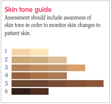
Figure 1.
 Skin tone tool (adapted from Ho and Robinson, 2015). 

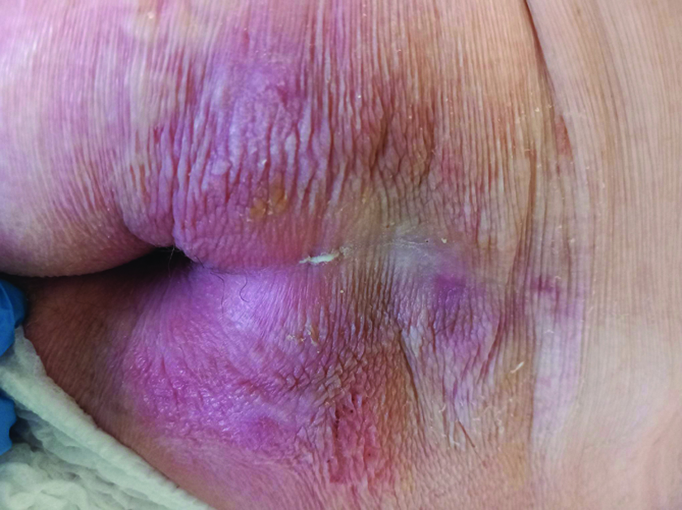
Figure 2.
Erythema is clearly visible in skin type 1.

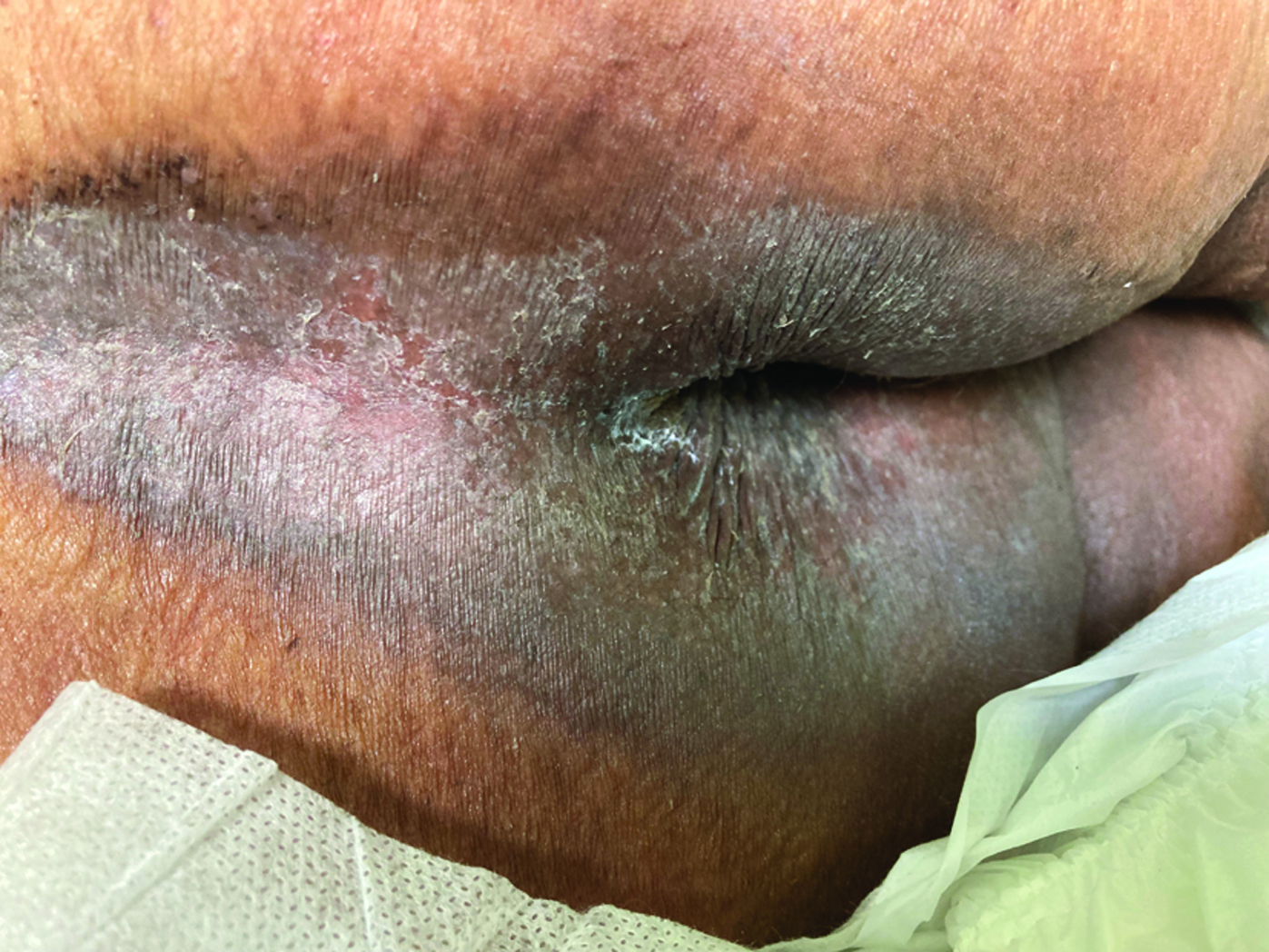
Figure 3.
Erythema is less obvious in skin type 4. 

SKIN TONE AND IAD ASSESSMENT  


In moderate-to-severe IAD, the epidermis breaks down and in light skin it will appear pink or red. The affected area usually has poorly defined edges and may be patchy or continuous over large areas (Beeckman et al, 2015).  

When identifying ‘redness’ in IAD, consideration must be given to how this may present in a range of skin tones, even if this is not always obvious in literature and guidelines. For example, the Ghent Global IAD Categorisation Tool (GLOBIAD) categorises IAD severity based on visual inspection of the affected skin areas (Beeckman et al, 2018). It relies on ‘redness’ as a key indication of damage and all images are of light skin tones. It does, however, acknowledge that a variety of tones of redness may be present; and in patients with darker skin tones, the skin may be paler or darker than normal, or purple rather than red (Beeckman et al, 2018).  

The unique characteristics of dark skin tones should be taken into account when assessing IAD. They include higher lipid content and more melanosomes in the stratum corneum compared with light skin. These characteristics may cause inflammation to present as violet-black or black, which may mask erythema. Because darker skin has more melanin than light skin, assessment of blanching can also be muted, complicating identification of early skin injury (Francis, 2019).  

It is worth noting that skin irritation in patients with dark skin tones may cause hyperpigmentation (increased pigmentation) or hypopigmentation (reduced pigmentation), with no redness visible (Nijhawan and Alexis, 2011).  

Changes in skin colouration are often the main visual sign of erythema in dark skin. This can be easier to identify when affected areas are compared with unaffected skin. Figures 2 and 3 demonstrate that while erythema is obvious in skin type 1 (Figure 2), it is less easy to diagnose in skin type 4 (Figure 3). Yet, the patient with skin type 4 is in the early stages of IAD. Without an early diagnosis, the skin is likely to breakdown, which may even lead to a pressure ulcer forming.  

IAD MANAGEMENT CONSIDERATIONS  


The skin should be examined thoroughly and regularly, taking into account skin tone. This should form part of an overall holistic assessment which includes not just the patient’s skin, but also their overall health and medical history, together with any significant cultural considerations (Wounds UK, 2021). Taking this approach means that care can be individually tailored to meet each patient’s needs. Regular skin assessments mean that any changes can be managed at an early stage, with protective measures such as barrier and cleansing products used before damage occurs.  

When caring for patients with IAD, it is important to remember that skin tone is separate from race. For example, not all people classified as black have dark skin tones; and not all those classified as white have light skin tones. In the author’s clinical opinion, this is where the skin tone assessment tool is invaluable, as it shows a range of six skin tones, not linked to ethnicity.  

It is important to acknowledge that different patient groups may have different needs. In some communities people may be mistrustful of the healthcare system and be reluctant to seek help (Mukwende, 2020). This reluctance has been intensified by the recent pandemic when access to health care was severely limited. Indeed, in the author’s clinical experience, many patients only seek clinical intervention for IAD when their symptoms have a major impact on their everyday life.  

When assessing a patient with IAD, it is always worth asking direct questions, such as, ‘are any parts of your skin sore?’ or ‘have you noticed any changes to your skin?’ This can help to obtain information that might otherwise have been missed.  

Selecting treatments and products tailored to the individual patient is essential. Wherever possible, shared decision-making should take place, so that the individual feels engaged with and confident in the care they are receiving (Moore et al, 2015). Supported self-care should be encouraged where possible, with education about the importance of skin integrity and protection forming part of the care plan (Fletcher et al, 2020). 

Skin protection products such as barrier creams should be used to create a protective layer on the skin’s surface, that simultaneously maintain hydration levels while blocking external moisture and irritants (Waller and Cole, 2023). Appropriate continence pads also have a significant role to play in management.  

The skin of patients who are incontinent should be cleansed at least once daily and after each episode of faecal incontinence (Beeckman et al, 2015). The author’s clinic (CWC) prefers cleansing products which are simple to use with good skin compatibility. Antimicrobial octenidine-impregnated wash mitts are regularly used for cleaning intact skin when managing IAD. The clinic’s qualitative data shows patient and professional satisfaction with the mitts (Dhoonmoon and Dyer, 2020). Octenidine is an antimicrobial with good tissue compatibility (Vanscheidt et al, 2012), and is known to prevent bacterial growth (Cutting and Westgate, 2012). It also has deodorising properties, which is an advantage when managing IAD. In the author’s clinical experience, the mitts are easy to use in a non-clinical setting, which can help improve compliance.  

PRACTICE POINTS 

  • ● Always listen to the patient’s perspective on their wound and overall health  
  • ● Use neutral and professional terminology which focuses on skin tone  
  • ● Inspect the skin thoroughly and regularly, so that diagnosis can be made before maceration occurs 
  • ● Incorporate the skin tone assessment tool into existing IAD management frameworks. 

CASE REPORTS  

Patient A  


Patient A was referred to the CWC by her general practice nurse (GPN) who was concerned about non-healing ‘sores’ on the patient’s buttocks, which were causing pain and intense itching. These symptoms were having a significant impact on her life. She was mobile and quite independent but did have a history of recurrent falls. The patient lives with her daughter who supports her with cooking, cleaning and food shopping.  

Her previous medical history included diverticular disease, gastric polyp, hiatus hernia, fracture of the humerus, hypertension and chronic obstructive pulmonary disease (COPD).  

At initial assessment, pressure areas and skin integrity were checked. Assessment included a skin tone evaluation, which was recorded as type 3. She had a broken moisture lesion to her buttocks and natal cleft; and there were clear signs of venous engorgement. It is likely that the early signs of erythema were missed by her carers, due to her skin tone making diagnosis less obvious.  

The patient advised that she ‘dribbles’ constantly, so wears a sanitary towel. She is changing this more frequently due to the amount of blood leaking from the moisture lesion. She also reported that she sweats heavily, particularly in the anal area, which has made the area feel very itchy. The patient’s urinary incontinence and perspiration contributed to the breakdown of her skin. The itchiness caused the patient to scratch her buttocks, causing more excoriation to the already vulnerable area.  

An enzyme alginogel was prescribed for use on the open wound, with octenidine-impregnated wash mitts to help manage irritation in the periwound areas. Her daughter was shown how to manage the skin care regimen and was fully engaged with the treatment plan. The patient declined referral for continence assessment, stating a preference to ‘manage this herself’.  

Eight weeks after initiating the treatment plan, the moisture lesions were considerably smaller and were causing little or no discomfort to the patient. At 12 weeks, there was evidence of complete healing. It was agreed that no further visits were required, and that the management regimen would be continued.  

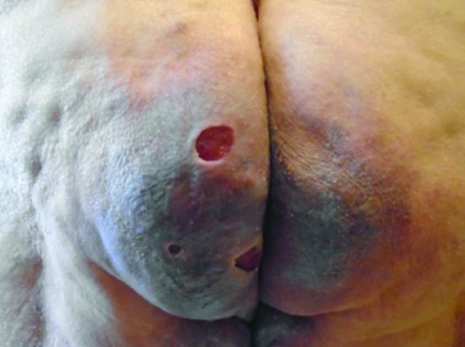
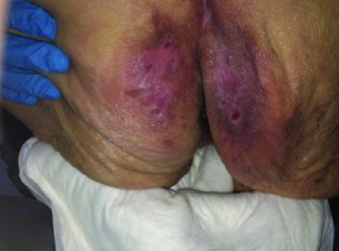
Patient A: at initial tissue viability assessment

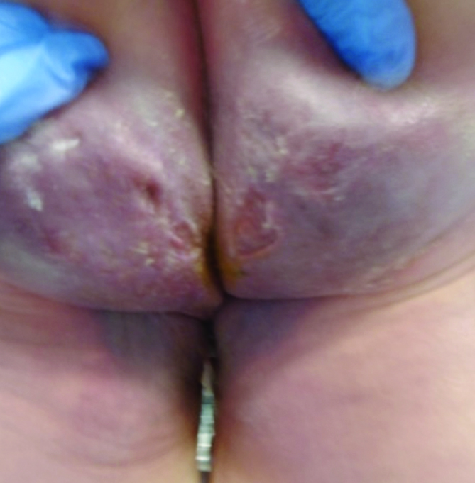
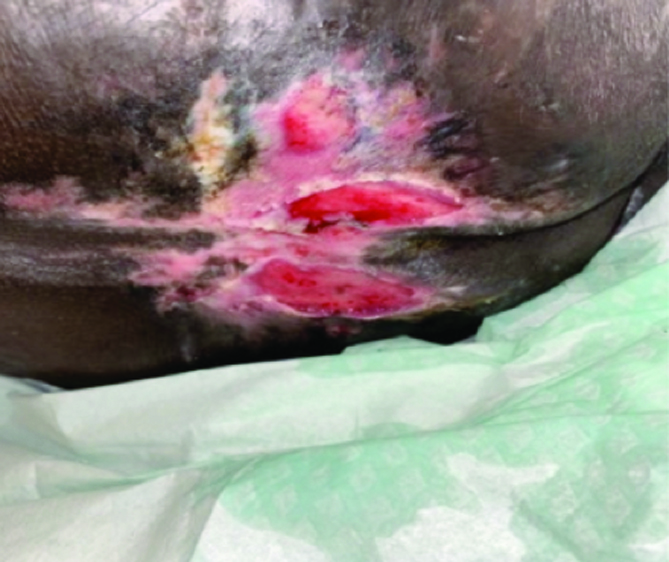
Patient A: four weeks later

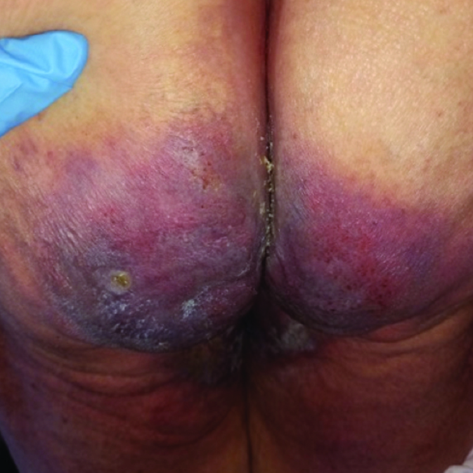
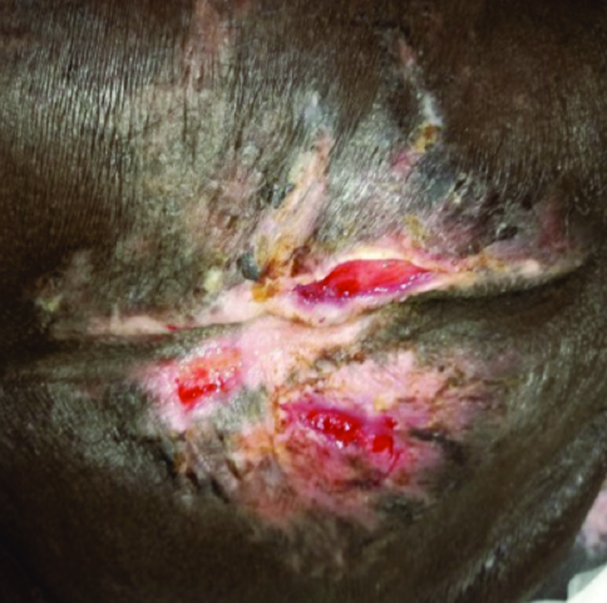
Patient A: eight weeks later

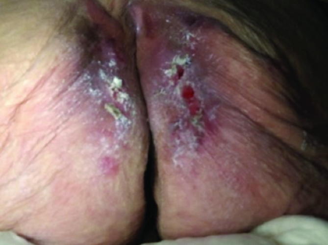
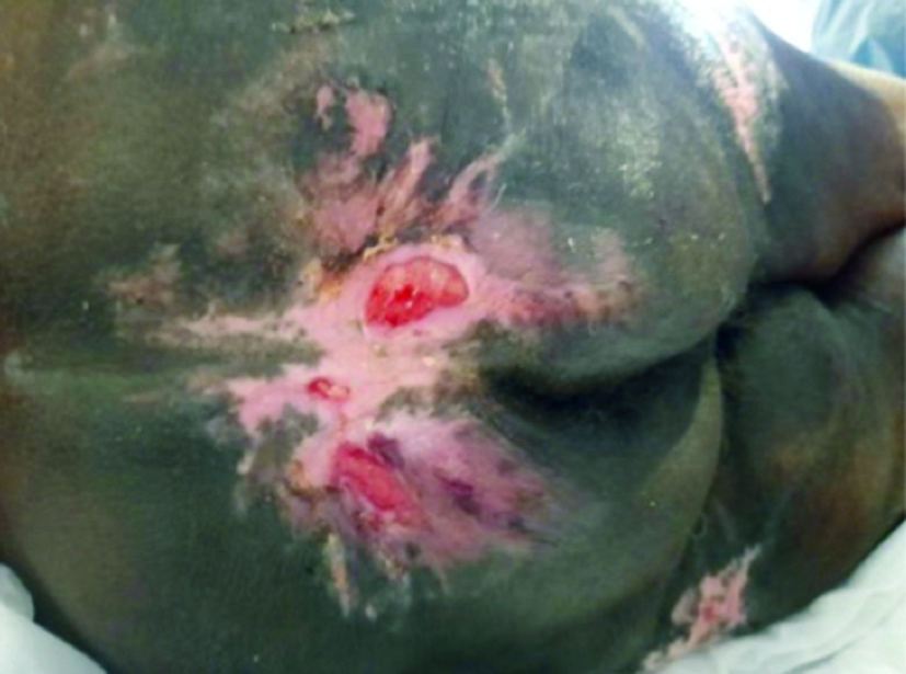
Patient A: 12 weeks later

Patient B  


Patient B was referred to the author’s clinic by her GP due to a chronic skin lesion to the buttocks.  

Her medical history included reduced mobility following a recent hip fracture, hypertension, raised cholesterol levels, type two diabetes mellitus and asthma She lives with her daughter who is her main carer and very supportive of her mother.  

At initial assessment, her skin was very dry and IAD was present. Her skin tone was type 2. After discussions with the patient, she stated that she had both faecal and urinary incontinence. The pain from the lesions was so severe that she could not sit down. Also, the odour from her skin disturbed her sleep and appetite. The patient’s treatment goal was to sit down to eat and enjoy a meal.  

Her daughter had been cleaning the buttock area regularly with scented bubble bath to help keep her mother ‘feeling clean and fresh’. Baby lotion was also applied to the area. Both products were likely to have increased skin irritation. Having skin tone type 2 and double incontinence meant that her daughter missed the early signs of IAD, only seeking medical intervention when there were obvious skin lesions.  

A management plan was discussed and agreed with the patient and her daughter, with good skin care being central to treatment. They agreed to discontinue the use of scented bubble bath and baby lotion. Instead, they were taught how to use octenisan wash mitts on the periwound areas, followed by a barrier cream. 

Four weeks later, the patient was able to sit out for lunch and evening tea with her daughter. No pain was reported, the lesions were healing, and odour was no longer an issue. No further clinical input was required, as they both felt well supported and wanted to continue the cleansing and barrier cream regimen. 

Patient B: at initial TV assessment

Patient B: four weeks later

Patient C  


Patient C was referred to the author’s clinic by the district nurse due to skin deterioration on both buttocks linked to double incontinence. She lives alone at home but is bedbound and supported by a four times daily package of care.  

Her medical history included advanced stage Alzheimer’s disease, faecal and urinary incontinence, hypertension, raised cholesterol and type 2 diabetes mellitus. She had been offered catheterisation to manage her urinary incontinence but declined this intervention, preferring to use pads.  

At initial assessment, the skin was eroded and burning pain was being caused by extensive excoriation. A diagnosis of IAD was made, and her skin tone was recorded as 6, meaning that the early stages of IAD had not been diagnosed by her carers.  

Although her carers had been using a barrier cream, the patient’s faecal incontinence caused the skin to continue breaking down, leading to further erosion. Due to pain, the patient did not allow carers to deliver personal care.  

The management plan included liaison with the local continence service to ensure that the patient had the correct pads; and with the occupational therapy team to install the required pressure-relieving equipment in her home.  

Carers were advised to clean faeces with plain wipes and warm water first, then use octenidine-impregnated wash mitts to remove any contaminants and bioburden from the intact areas of skin. A skin protectant ointment was applied to the lesions. 

At the four-week review with the patient and her carers, they reported that the patient accepted this management plan as the cleansing regimen did not cause pain. She was much calmer and not shouting or crying when approached for personal care. They also noted that the patient’s appetite had improved. Eight weeks later, further healing was noted. At 12 weeks, her skin integrity showed significant improvement and as healing continued to progress, she was discharged from the care of the tissue viability team. 

Patient C: at initial TV assessment

Patient C: eight weeks later

Patient C: 12 weeks later

CONCLUSION  


All forms of MASD, including IAD, have a significant impact on patient wellbeing and quality of life, so it is vital that all care is delivered using best practice and with a patientcentred approach. Optimising treatment and outcomes depends on accurate skin assessment, for which knowledge of the signs and symptoms across skin tones is essential (Mukwende, 2020). This means that training is needed so that all clinicians and carers are confident to make this assessment and understand its importance in best practice. Healthcare professionals have a responsibility to prevent health inequality, so recognising and taking into account skin tones is an opportunity to improve care. This will help to create an environment in which patients feel confident in the care they receive, regardless of their skin tone.  
Luxmi Dhoonmoon is nurse consultant tissue viability, London North West University Healthcare NHS Trust. 
This piece was first published in the Journal of General Practice Nursing. To cite this article use: Dhoonmoon L (2023) Skin tone in the management of incontinence-associated dermatitis. J Gen Practice Nurs 9(3): 34–40 

References

Beeckman D, Campbell J, Campbell K, et al (2015) Proceedings of the Global IAD Expert Panel. Incontinence Associated Dermatitis: Moving Prevention Forward. Wounds International, London  

Beeckman D, Van den Bussche K, Alves P, et al (2018) Towards an international language for incontinence-associated dermatitis (IAD): design and evaluation of psychometric properties of the Ghent Global IAD Categorization Tool (GLOBIAD) in 30 countries. Br J Dermatol 178(6): 1331–40  

Cutting K, Westgate SJ (2012) The use of wound cleansing solutions in chronic wounds. Wounds UK 8: 130–3 

Dhoonmoon L, Dyer M (2020) Improving leg ulcer care in the community. J Community Nurs 34(6): 40–4 

Fletcher J, Beeckman D, Boyles A (2020) International Best Practice Recommendations. Prevention and management of moistureassociated skin damage (MASD). Wounds International. Available online: www.woundsinternationalcom   

Francis K (2019) Damage control: Differentiating incontinence-associated dermatitis from pressure injury. Nursing 14(6): 18–25  

Glass GF Jr, Goh CCK, Cheong RQ, Ong ZL, Khong PCB, Chan EY (2021) Effectiveness of skin cleanser and protectant regimen on incontinenceassociated dermatitis outcomes in acute care patients: A cluster randomised trial. Int Wound J 18(6): 862–73 (published correction appears in Int Wound J [2022] 19(7): 1955)  

Gov.UK (2021) Ethnicity facts and figures. Available online: www.ethnicity-factsfigures.service.gov.uk/   

Gray M, Black JM, Baharestani MM, et al (2011) Moisture-associated skin damage: overview and pathophysiology. J Wound Ostomy Continence Nurs 38(3): 233–41  

Green M, Kashetsky N, Feschuk A, Maibach HI (2022) Transepidermal water loss (TEWL): environment and pollution - A systematic review. Skin Health Dis 2(2): e104  

Gupta V, Sharma VK (2019) Skin typing: Fitzpatrick grading and others. Clin Dermatol 37(5): 430–6  

Ho BK, Robinson JK (2015) Color bar tool for skin type self-identification: A crosssectional study. J Am Acad Dermatol 73(2): 312–3  

Lopez-Ojeda W, Pandey A, Alhajj M, et al (2022) Anatomy, Skin (Integument) [Updated 2022 Oct 17]. In: StatPearls [Internet]. Treasure Island (FL): StatPearls Publishing; 2023 Jan. Available online: www.ncbi.nlm.nih.gov/books/NBK441980/   

Meyer I, Richter HE (2015) Impact of fecal incontinence and its treatment on quality of life in women. Womens Health (Lond) 11(2): 225–38  

Moore Z, Bell T, Carville K, et al (2015) International Best Practice Statement: Optimising patient involvement in wound management. Wounds International, London  

Mukwende M (2020) Mind the Gap: a clinical handbook of signs and symptoms in black and brown skin. Wounds UK 16(3): 16  

Nakagami G, Sanada H, Kitagawa A, et al (2006) Incontinence induces stratum corneum vulnerability and impairs the skin barrier function in the perianal region. Dermatology 213(4): 293–9  

NHS England (2018) Excellence in Continence Care. Available online: www.england.nhs.uk/wp-content/uploads/2018/07/excellence-in-continence-care.pdf    

Nijhawan R, Alexis A (2011) Practical approaches to medical and cosmetic dermatology in skin of color patients. Expert Rev Dermatol 6: 175–87  

Norton C, Thomas L, Hill J (2007) Guideline Development Group. Management of faecal incontinence in adults: summary of NICE guidance. BMJ 334(7608): 1370–71  

Vanscheidt W, Harding K, Teot L, Siebert J (2012) Effectiveness and tissue compatibility of a 12-week treatment of chronic venous leg ulcers with an octenidine based antiseptic — a randomized, double-blind controlled study. Int Wound J 9(3): 316–23  

Waller S, Cole N (2023) Versatility of a skin protectant ointment in managing moisture-associated skin damage caused by incontinence. Wounds UK 19(1): 58–63  

Woo KY, Beeckman D, Chakravarthy D (2017) Management of moistureassociated skin damage: a scoping review. Adv Skin Wound care 30(11): 494–501 

 Wounds UK (2021) Best Practice Statement: Addressing skin tone bias in wound care: assessing signs and symptoms in people with dark skin tones. Wounds UK, London. Available online: www.wounds-uk.com 

Woodward S, Norton C (2020) New study into treatment and prevention of incontinence-associated dermatitis. King’s College, London. Available online: www.kcl.ac.uk/news/new-studyinto-treatment-and-prevention-ofincontinence-associated-dermatitis   

Young T (2017) Back to basics: understanding moisture-associated skin damage. Wounds UK 13(2): 56–65