Introduction

The skin is an important organ of the body which performs a number of key functions that are vital to our overall health and wellbeing. These are sometimes referred to as internal haemostasis or the homeostatic functions of the skin (Wingfield, 2011; Dowsett and Allen, 2013). Intact healthy skin has a pH of around 5 (Penzer, 2012) which allows it to function optimally as a barrier to the outside world. However, when the pH is disrupted, the barrier function of the skin is compromised, leading to skin damage and a proliferation of bacteria (Beeckman et al, 2015). This article will describe the structure and function of the skin, how normal pH is maintained, factors that disrupt it and the consequences of this on skin function and provide guidance on how to restore skin pH.
 

Structure of the skin

The skin consists of two layers, the epidermis and dermis, that are divided by the basement membrane which provides nutrients and strength to the epidermis (Watkins, 2013).


Epidermis

The epidermis is avascular and entirely dependent on the dermis below (Lawton, 2019). It is made up of five layers and each layer represents the different maturity of the keratinocyte, the predominant cell of the epidermis which produces keratin and forms the epidermal water barrier (Watkins, 2013). The other cells are; melanocytes which produce melanin and offer protection from the damaging effects of radiation from the sun, Langerhans’ cells which play a part in immunity, and Merkel cells which are integral to touch sensation (Watkins, 2013) (figure 1).

The innermost layer is the stratum basale layer, a single layer of cells that produce all of the cells of the epidermis and which gradually migrate upwards (Wheeler, 2012).

The outer layer, the stratum corneum, is the most important layer in maintaining the pH-dependent barrier function of the skin, essential for preventing infection and minimising water loss (Schmid-Wendtner and Korting, 2006; Wheeler, 2012; Jones, 2014).

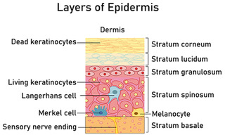
Figure 1 - The layers of the epidermis.

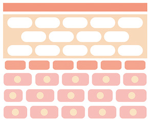
Figure 2 - Model of the stratum corneum
The stratum corneum is made up of 15–20 layers of fully cornified keratinocytes – corneocytes (Beeckman et al, 2015; Boer et al, 2016). In the bottom part of the cornified layer, the cells are closely joined to each other with desmosomes (Beeckman et al, 2015; Boer et al, 2016). In the top part of the layers they are arranged more loosely and undergo scaling and shedding (Boer et al, 2016). The corneocytes are embedded in intercellular lipids which are often described as bricks and mortar where the bricks are the corneocytes and the intercellular lipids are the mortar (Beeckman et al, 2015; Boer et al, 2016) (Figure 2). The corneocytes contain a natural moistening factor which provides hydration for effective skin function (Beeckman et al, 2015). The size of the corneocytes and the composition of the lipids impact the regeneration properties of the skin (Boer et al, 2016) which is important when considering skin barrier products which are formulated with lipids similar to those found in healthy stratum corneum, reducing skin dryness and restoring the lipid matrix (Beeckman et al, 2015). Exposure to urine and/or faeces and repeated washing with an alkaline soap changes the pH of the skin and eventually breaks down the intracellular lipid layer mortar and the natural moisturising factor within the corneocytes, breaking down the barrier within the stratum corneum and making it vulnerable to further damage (Proksch, 2018).   
 

Dermis

The dermis is a thicker, deeper layer predominantly made up of fibrous proteins, collagen and elastin which gives the skin its strength and elasticity (Jones, 2014). It contains hair follicles, sebaceous glands (sebum), apocrine glands (scent), eccrine glands (sweat), blood vessels and nerves (Watkins, 2013).
 

Hypodermis

Below the dermis is the subcutaneous layer also known as the hypodermis which is made up of adipose tissue and connective tissue (Jones, 2014) and contains larger blood vessels and nerves and is responsible for the regulation of temperature and acts as a shock absorber (Watkins, 2013). 

All three layers combine together to perform several functions, but the epidermis is the most important layer that protects the inner body from the environment (Peters, 2008).
 

Skin function

The skin has many important functions (Table 1) but the main function of the skin is to provide a protective barrier (Penzer, 2012). The condition of the epidermal barrier depends on its physical properties, such as the amount of sebum produced, epidermis hydration, transepidermal loss of water and the pH gradient between the skin surface and the inside of the body (Boer et al, 2016). When intact, the skin prevents the penetration of pathogens and allergens through its surface (Wounds UK, 2018). This barrier also plays a part in ensuring that water is held in the skin (Penzer, 2012). The barrier is enhanced by the acid mantle, which refers to the surface of the skin being slightly acidic (Wingfield, 2011).
Table 1 – Other important functions of the skin
  1. Protection - the skin produces melanin which is responsible for skin colouring and protection against ultraviolet light. The skin also forms a barrier against trauma, protecting the internal tissues and structures from damage (Wounds UK, 2012)
  2. Sensory perception – where the nerve endings respond to pain, temperature, vibration and touch (Wounds UK, 2018)
  3. Temperature regulation – vasodilation to cool the body and vasoconstriction to retain heat (Wounds UK, 2012) along with the production of sweat (Penzer, 2012)
  4. Production of vitamin D – which regulates calcium and phosphate supplies in body fluids (Wingfield, 2011).
How normal pH is maintained  
A major part of the skin’s ability to provide a protective barrier is maintaining a constant pH (denoting the power or potential of hydrogen) of between 4 and 6 (Beeckman et al, 2015). A pH of 7 is considered neutral (water) anything below is acidic and anything above alkaline (Figure 3).

This slightly acidic nature of the skin, known as the acid mantle, is an antimicrobial barrier and a barrier against permeability (Wounds UK, 2018).

The formation of the stratum corneum barrier involves several pH-dependent enzymes, such as β-glucocerebrosidase, acid lipases and phosphatases. β-glucocerebrosidase with an optimum pH of 5.6 is involved in the synthesis of the most important lipids (fat molecules) that help the skin retain moisture and allow for proper function (Schmid-Wendtner and Korting, 2006).
 
 
Figure 3 The pH scale.

Factors affecting skin pH

Skin pH can be influenced by a number of internal/physiological and external factors including:
  • Anatomical site – there are slight variations in the skin’s pH between the face, trunk and extremities. The pH in physiological gaps, such as the axilla and groin, are between 6.1 and 7.4 giving them a different commensal microbial community adapted to the pH of the local environment. Transfer of micro-organisms from one location to another can therefore result in infection (Proksch, 2018).
  • Age – the structure and function of the skin’s barrier deteriorates with the ageing process and so as the skin becomes thinner it also becomes more vulnerable to damage, injury and dryness (Wingfield, 2011). The stratum corneum pH is higher in newborns than older children because of the incomplete acidification (Schmid-Wendtner and Korting, 2006; Proksch, 2018)
  • Ethnic differences – darkly pigmented skin exhibits a lower pH (4.6 rather than 5). There also appears to be an increased epidermal lipid content giving a better skin surface integrity and barrier function (Proksch, 2018)  
  • Soaps and cosmetic products - some soaps and cosmetic products can be alkaline and can change the delicate pH balance of the skin (Proksch, 2018; Wounds UK, 2018) and some are irritants to the skin (Penzer, 2012)
  • The presence of urine and faeces on the skin – urine and/or faeces can cause swelling of the stratum corneum and change the pH to alkaline resulting in incontinence-associated dermatitis (IAD) (Beeckman et al, 2015).

Consequences of IAD on the skin barrier function

The condition where urine and faeces damages the skin is known as IAD. IAD is mainly a chemical irritation, similar to contact dermatitis, when urine or faeces comes into contact with the skin (Dowsett and Allen, 2013; Young, 2017; McNichol et al, 2018). The lipid- and protein-digesting enzymes from faeces, combined with ammonia produced by the conversion of urea found in urine, increase the skin’s pH to alkaline which destroys the barrier mechanism by decreasing stratum corneum cohesion and allows bacteria to thrive (Beeckman et al, 2015; Beele et al, 2018). Enzymes are more active with a higher pH and so combined urine and faecal incontinence is more damaging (Beeckman et al, 2015). Liquid faeces has the highest number of digesting enzymes and can be the most damaging (Beeckman et al, 2015).   
pH of the skin is important for antimicrobial activity and acids are known for their role in inhibiting the colonisation of bacteria (Proksch, 2018). The growth pace and the colonisation density of bacteria and fungi increase with the increase of pH (Boer et al, 2016). Non-pathogenic skin flora normally reside on the skin and adapt to the pH, but an acid pH inhibits the colonisation of harmful, pathogenic bacteria (Boer et al, 2016; Proksch, 2018).  

Washing with alkaline soaps result in an increased pH allowing bacteria to grow (Beeckman et al, 2015; Proksch, 2018). It has been shown that a pH rise lasts for several hours after cleaning the skin with alkaline soap (Schmid-Wendtner and Korting, 2006) which means the risk of pathogenic bacteria colonisation is high until the pH is restored.

Maceration also plays a key role in damaging the skin barrier (Dowsett and Allen, 2013). Water from urine and faeces is pulled into and retained by corneocytes which causes swelling and disrupts the structure of the stratum corneum, increasing the skin-support coefficient of friction and allowing irritants to enter the skin (Beeckman et al, 2015; Beele et al, 2018). Some containment products have been specifically designed to absorb fluids that can cause skin damage and contain fibres that are chemically designed to maintain the skin’s pH (Rippon et al, 2016). These newer, improved products ensure that special fibres are contained within the upper layer of the pad material and sit directly next to the skin to buffer the pH (e.g. MoliCare® range of pads and underwear by HARTMANN).   
IAD can occur with faecal incontinence, double incontinence and urinary incontinence with the risk increasing greatly with liquid faeces because of the higher level of damaging enzymes (Beeckman et al, 2015). Product selection is key to minimising the risk of IAD and to ensure newer products are used with improved fluid handling properties minimising the contact of moisture with the skin, as an adjunct to a structured skin regimen (Beeckman et al, 2015).  

Other key risk factors for IAD that relate to the skin include:
  • Poor skin condition – e.g. due to the ageing process, use of steroids or conditions such as diabetes
  • Poor nutrition – good nutrition is vital for skin health and maintenance of the barrier function. Protein is required for keratin production, vitamins and minerals for skin regeneration and fatty acids for skin renewal and hydration
  • Occlusive containment products – products with poor absorbent properties hold moisture against the skin (Beeckman et al, 2015; Wounds UK, 2018).     
It is important that clinicians know and can recognise the difference between IAD and pressure ulcers as there has long been confusion (Ousey and O’Connor, 2017; McNichol et al, 2018). IAD has been described as a ‘top down’ injury, beginning on the surface of the skin whereas pressure ulcers are seen as a ‘bottom up’ injury with the damage starting below the skin in the soft tissues (Beeckman et al, 2015; Ousey and O’Connor, 2017).

The use of an IAD severity tool is recommended in best practice (Beeckman et al, 2015; Beeckman, 2017; Young, 2017) and is described in box 1.
Box 1 IAD severity tool (adapted from Beeckman et al, 2015; Young, 2017)
Severity  Signs 
At risk Skin intact/no redness  Skin normal when compared to rest of body  
Mild to moderate
Category 1A – Persistent redness without infection (a variety of colours may be seen in darker skin tones)
Category 1B – Persistent redness with infection  
Erythema
Swollen or tense skin
Shiny appearance
Macerated skin
Pain/burning/itching
Intact vesicles and bullae  
Moderate to severe
Category 2A – Skin breakdown without infection
Category 2B – skin breakdown with infection  
Erythema (a variety of colours may be seen in darker skin tones)
Swollen or tense skin
Shiny appearance
Macerated skin
Pain/burning/itching
Intact vesicles and bullae  
Restoring skin pH with a structured skin care routine
Best practice guidance recommends that a structured skin care routine is based on the acronym CPR (cleanse, protect and restore) in order to maintain the skin’s pH (Beeckman et al, 2015; Young, 2017).


Cleanse

The dehydrating effects of soap on the natural lipids in the skin has been known for some time (Peters, 2008; Ousey and O’Connor, 2017). Soap also tends to be alkaline which further disturbs the acid mantle and the balance of the skin pH (Peters, 2008). Friction caused by over-vigorous use of washcloths and rubbing is also known to be detrimental to the skin (Beeckman et al, 2015). Cleansing should take place as soon as possible after an episode of incontinence (Ousey and O’Connor, 2017). A pH balanced, no-rinse cleanser (e.g. MoliCare® Skin Cleanse Foam) specifically for use in incontinence should be used (Beekman et al, 2015) as it reduces skin irritation and dryness reducing the risk of compromising skin integrity problems (McNichol et al, 2018).  
 

Protect

The principles of protection are to provide a barrier between the skin and urine and/or faeces and to retain the moisture content in the skin (Ousey and O’Connor, 2017; McNichol et al, 2018). This can be achieved with a moisturiser in people at risk of, or with, mild IAD (Young, 2017). A specifically designed skin protectant can be used for more severe cases of IAD (Beeckman et al, 2015) as they allow the skin to recover. It is important to select a barrier product that does not affect the absorbency of containment products (e.g. MoliCare® Skin care products) and avoid those that may affect the way fluid is taken into the absorbent core (e.g. petroleum jelly) (Beeckman et al, 2015).
 

Restore

Some patients may have the need for an extra step in their skin care to help restore the skin’s barrier properties and this usually involves the application of products that are left on the skin. Such products are usually reserved for severe cases, or patients at very high risk, of IAD (Beeckman et al, 2015; Young, 2017).

A Cochrane review found the use of leave-on products such as moisturisers, skin protectants, or a combination of both, and avoiding soap use, was more effective for preventing and treating IAD in adults than not using these products (Beeckman et al, 2016).

A skin care regimen for the treatment of IAD may include the use of separate products to cleanse and protect (Beeckman et al, 2015). For the prevention of IAD restoring the skin’s barrier function is desirable and so by using products that have a combined affect, e.g. protect and restore, it may be possible to reduce the steps involved in care and so save time in delivery of skin care (Beeckman et al, 2015).
 

Nutrition and hydration

Good nutrition is an important component of maintaining the skin barrier function, skin integrity and health (Collier and Simon, 2016; Wounds UK, 2018). Maintaining hydration is also important as dehydration can cause the skin to lose elasticity, making it more susceptible to damage  (Collier and Simon, 2016).


Conclusions

The skin has two layers, but it is the outmost layer that plays the biggest role in maintaining protective barrier function and preserving the delicate pH balance of the skin. Exposure of the skin to moisture, such as urine and faeces, has a very damaging effect on function. Therefore, in patients who suffer from incontinence, it must be proactively prevented and managed. One of the main ways to do this is to use a structured skin care routine to help cleanse, protect and restore the skin and maintain its pH.
 

References

Beeckman D, Campbell J, Campbell K et al (2015) Proceedings of the Global IAD Expert Panel. Incontinence-associated dermatitis: moving prevention forward. Wounds International, London. Available online: www.woundsinternational.com 

Beeckman D, Van Damme N, Schoonhoven L, et al (2016) Interventions for preventing and treating incontinence-associated dermatitis in adults. Cochrane Database of Systematic Reviews, Issue 11. Art. No.: CD011627. DOI: 10.1002/14651858.CD011627.pub2   

Beeckman D (2017) A decade of research on incontinence-associated dermatitis (IAD): Evidence, knowledge gaps and next steps. J Tissue Viability 26(1): 47-56

Beele H, Smet S, Van Damme N, Beeckman D (2018) Incontinence-associated dermatitis: pathogenesis, contributing factors, prevention and management options. Drugs Aging 35: 1-10

Boer M, Duchnik E, Maleszka R, Marchlewicz M (2016) Structural and biophysical characteristics of human skin in maintaining proper epidermal barrier function. Adv Dermatol Allergol 33(1): 1-5  

Collier M, Simon D (2016) Protecting vulnerable skin from moisture-associated skin damage. Br J Nurs 25(20): S26-S32

Dowsett C and Allen L (2013) Moisture-associated skin damage made easy. Wounds UK 9(4): 1-4. Available online: www.wounds-uk.com

Jones ML (2014) Treating vulnerable skin: the cornerstone of providing good care. Nurs Residential Care 16(12): 671-676

McNichol LL, Ayello EA, Phearman et al (2018) Incontinence-associated dermatitis: state of the science and knowledge. Adv Skin Wound Care 31(11): 502-513

Ousey K and O’Connor L (2017) IAD made easy. Wounds UK 13(1): 1-6. Available online: www.wounds-uk.com

Penzer R (2012) Best practice in emollient therapy: A statement for healthcare professionals. Dermatol Nurs 11(4): S1-S19

Peters J (2008) Managing eczema at either end of the age spectrum. Dermatol Nurs 7(2): S7-S11

Proksch E (2018) pH in nature, humans and skin.  J Dermatol 45: 1044-1052

Rippon M, Colegrave M, Ousey K (2016) Incontinence-associated dermatitis: reducing adverse events. Br J Nurs 25(18): 1016-1021

Schmid-Wendtner MH, Korting HC (2006) The pH of the skin surface and its impact on the barrier fiction. Skin Pharmacol Physiol 19: 296-302

Watkins J (2013) Skin rashes, part 1: Skin structure and taking a dermatological history. Pract Nurs 24(1): 30-33

Wheeler T (2012) Our outermost boundary: the skin. Nurs Residential Care 14(11): 570-576

Wingfield C (2011) Skin care in the older person: a focus on the use of emollients. Br J Community Nurs 16(10): 470-478

Wounds UK (2012) Best Practice Statement: Care of the older person’s skin. Available online: www.wounds-uk.com

Wounds UK (2018) Best Practice Statement: Maintaining skin integrity. Available online: www.wounds-uk.com  

Young T (2017) Back to basics: understanding Moisture-associated skin damage. Wounds UK 13(2): 56-65