The urinary tract is divided into:
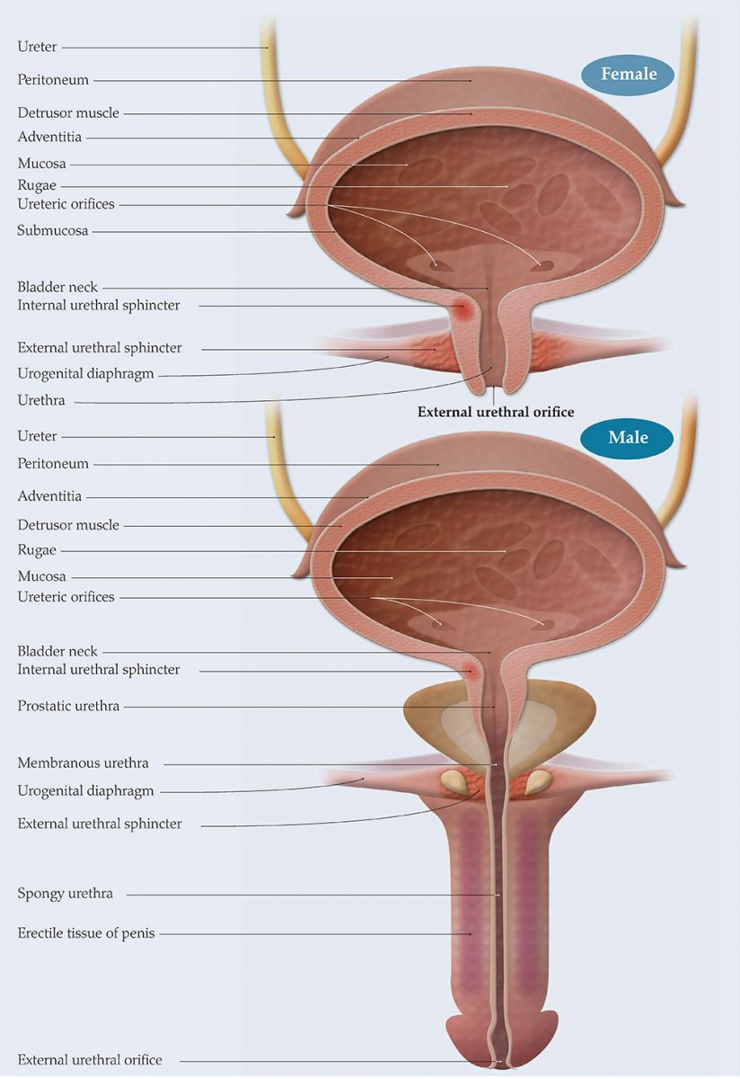
The bladder has four layers, the outermost of which is continuous with the peritoneum. The innermost layer is the internal mucosa made of transitional epithelium. This is surrounded by a muscle layer made up of circular and longitudinal muscles forming a sort of net around the bladder. The mucosal layer is connected to this muscle layer by the lamina propria, a connective-tissue layer. The bladder muscle layer is called the detrusor muscle. It is studded with stretch receptors and as the bladder fills with urine and stretches to accommodate it, nerve impulses relay the message via the sacral spine up the vertebral column to the higher micturition centres in the brain, i.e. the pons, and then to the cerebral cortex (Fillingham and Douglas, 2004).
The micturition centres in the brain and sacral spine must all be functioning and able to communicate with each other via the nerves connecting them for the bladder to fill and empty normally.
The normal adult bladder usually holds between 300–600ml of urine and this empties out via the urethra. An internal sphincter made of smooth muscle lies at the base of the bladder at the bladder neck. There is also an external sphincter, consisting of striated muscle which is lower down the urethra and in close contact with the pelvic floor. Both are closed while the bladder is filling, but open when the bladder needs to be emptied (Urology textbook, 2018).
The male urethra is between 15–22cm long, while the female urethra is only 3–5cm in length. The male urethra extends from the bladder neck, through the prostate gland and the pelvic floor, then down to the external urethral opening.
The female urethra extends from the bladder neck and through the pelvic floor to the external opening and shares a wall with the vagina, which is situated behind it (Fillingham and Douglas, 2004).
Meanwhile, the pelvic floor moves downwards allowing ‘funnelling’ of the bladder, which means it is in the best position to empty (Fillingham and Douglas, 2004). At the same time, nerve impulses send messages from the cerebral cortex via the pons and down the spine to the bladder, allowing relaxation of the internal sphincter and contraction of the bladder muscle. The muscle squeezing the bladder causes a rise in bladder pressure, which pushes the urine out past the open bladder neck, down the urethra, past the open external sphincter and then the rest of the urethra and out of the body. When empty, both sphincters close again, the pelvic floor moves back up and the bladder relaxes allowing it to continue to fill. Again, to fill and empty normally, the bladder muscle and sphincters need to coordinate when they open and close with muscle contraction and relaxation. This means that the nerves which supply the bladder, sphincters and pelvic floor must all be functioning properly (Royal College of Nursing [RCN], 2012).
Anything which interrupts this coordination, for whatever reason, will cause the patient to have filling and voiding problems, which may lead to incomplete emptying or incontinence.
- The upper tract, which includes the kidneys and ureters
- The lower tract, which consists of the bladder and urethra.
The bladder has four layers, the outermost of which is continuous with the peritoneum. The innermost layer is the internal mucosa made of transitional epithelium. This is surrounded by a muscle layer made up of circular and longitudinal muscles forming a sort of net around the bladder. The mucosal layer is connected to this muscle layer by the lamina propria, a connective-tissue layer. The bladder muscle layer is called the detrusor muscle. It is studded with stretch receptors and as the bladder fills with urine and stretches to accommodate it, nerve impulses relay the message via the sacral spine up the vertebral column to the higher micturition centres in the brain, i.e. the pons, and then to the cerebral cortex (Fillingham and Douglas, 2004).
The micturition centres in the brain and sacral spine must all be functioning and able to communicate with each other via the nerves connecting them for the bladder to fill and empty normally.
The normal adult bladder usually holds between 300–600ml of urine and this empties out via the urethra. An internal sphincter made of smooth muscle lies at the base of the bladder at the bladder neck. There is also an external sphincter, consisting of striated muscle which is lower down the urethra and in close contact with the pelvic floor. Both are closed while the bladder is filling, but open when the bladder needs to be emptied (Urology textbook, 2018).
The male urethra is between 15–22cm long, while the female urethra is only 3–5cm in length. The male urethra extends from the bladder neck, through the prostate gland and the pelvic floor, then down to the external urethral opening.
The female urethra extends from the bladder neck and through the pelvic floor to the external opening and shares a wall with the vagina, which is situated behind it (Fillingham and Douglas, 2004).
EMPTYING THE BLADDER
The sensation of fullness when the bladder fills with urine triggers off a response, which alerts that it needs to be emptied. For this to happen, both sphincters need to relax and open while the bladder muscle contracts and expels the urine out of the body via the urethra. Unlike the internal sphincter muscle, which is not under voluntary control, the external sphincter can be controlled, and when it is time to void, this muscle relaxes to allow it to open.Meanwhile, the pelvic floor moves downwards allowing ‘funnelling’ of the bladder, which means it is in the best position to empty (Fillingham and Douglas, 2004). At the same time, nerve impulses send messages from the cerebral cortex via the pons and down the spine to the bladder, allowing relaxation of the internal sphincter and contraction of the bladder muscle. The muscle squeezing the bladder causes a rise in bladder pressure, which pushes the urine out past the open bladder neck, down the urethra, past the open external sphincter and then the rest of the urethra and out of the body. When empty, both sphincters close again, the pelvic floor moves back up and the bladder relaxes allowing it to continue to fill. Again, to fill and empty normally, the bladder muscle and sphincters need to coordinate when they open and close with muscle contraction and relaxation. This means that the nerves which supply the bladder, sphincters and pelvic floor must all be functioning properly (Royal College of Nursing [RCN], 2012).
Anything which interrupts this coordination, for whatever reason, will cause the patient to have filling and voiding problems, which may lead to incomplete emptying or incontinence.
References
Fillingham S, Douglas J, eds (2004) Urological Nursing. 3rd edn. Bailliere Tindall, UK
Royal College of Nursing (2012) Catheter care RCN guidance for nurses. RCN, London. Available online: http:// studyres.com/doc/8027570/cathetercare- rcn-guidance-for-nurses?page=2
Urology textbook (2018) Anatomy of the Bladder. Available online: www.urologytextbook. com/bladder-anatomy.html (last accessed 30 January, 2018)


