Faecal incontinence is the term describing the inability to control the bowels (International Continence Society [ICS], 2015). This can include the uncontrolled passage of solid or liquid stool, or flatus (wind) (Benezech et al, 2016) through the anal canal. It is recognised as a symptom rather than a diagnosis (National Institute for Health and Care Excellence [NICE], 2014) and is the result of complex interactions of many contributing factors. This series of two articles will look at this taboo condition, how it affects quality of life, highlight the anatomy and physiology which affects bowel control and then discuss conditions and contributing factors that make individuals prone to the symptoms of faecal incontinence. 

Faecal incontinence (FI) is one of the final taboo conditions that presents in health care today. Although common, it is a topic that is unlikely to be discussed by individuals in normal daily conversation due to embarrassment, shame and feelings of degradation. It is also a topic that is avoided or often forgotten to be asked about by healthcare professionals, meaning that most individuals with FI will suffer in silence for many years before discussing with family or professionals (Young, 2022). Faecal incontinence has been shown to have serious consequences on individuals, affecting their self-esteem and body image and creating feelings of shame and embarrassment, which can impact their social needs (International Continence Society [ICS], 2015; Camilleri-Brennan, 2020).  

FI is a devastating condition, which can result in social isolation, depression, skin breakdown and pose a financial burden (i.e. laundry costs, purchase of management equipment, e.g. protective pads, etc). It is also associated with secondary morbidities and disabilities and has a severe negative effect on an individual’s quality of life (ICS, 2015). Studies show it can lead to repeated urinary tract infections (UTIs) and premature admission to a care home (Potter et al, 2007; Harari, 2009; Norton et al, 2010). Furthermore, Duelund-Jakobsen et al (2016) state that individuals with FI have reduced ability to work and that it impacts their sexuality as they worry about smells and accidents during intimacy. Indeed, individuals will go to extreme lengths, spend considerable time, effort and attention avoiding situations where these unpredictable accidents may occur (Duelund-Jakobsen et al, 2016). 

PREVALENCE AND DEFINITION  


It is surprisingly common and is the term describing the inability to control the bowels (ICS, 2015). It is considered to be underreported due to the shame and embarrassment associated with it (Sbeit et al, 2021), and affects women, men and children. Prevalence figures estimate that approximately 0.5–1% of adults regularly suffer with FI (Royal College of Nursing [RCN], 2019). However, it is closely associated with age — over 15% of over 85 year olds living at home have FI and this increases to 10–60% of those living in residential or nursing home care (RCN, 2019). FI is a condition that is often underreported due to various factors, such as social stigma, embarrassment, and lack of awareness, and so measurement of actual prevalence and incidents are subjective and rely on patient reports. 
According to Benezech et al (2016), faecal incontinence is defined as the involuntary loss of flatus (wind) and/or solid or liquid stool, through the anal canal and the inability to postpone an evacuation until socially convenient. Attached to this definition is also a time duration of having the problem for at least one month and an age component of being at least four years old with previously achieved control (Benezech et al, 2016). The RCN (2019) further refined these definitions to include FI as the:   
  • Involuntary loss of liquid or solid stool that is a social or hygienic problem 
  • Anal incontinence (AI) as the involuntary loss of flatus, liquid or solid stool which is a social or hygienic problem 
  • Passive soiling (liquid or solid), which occurs when an individual is unaware of liquid or solid stool leaking from the anus; this may be after a bowel movement, or at any time  
  • Some definitions also include urge faecal incontinence whereby contents of the bowel are discharged despite active attempts to retain contents 
  • And faecal seepage (leakage of stool with grossly normal continence and evacuation).   
Assmann et al (2022) state it is defined as ‘The recurrent uncontrolled passage of faecal material for at least 3 months’, as per Rome IV criteria. Based on the frequency and quantity of FI, it can further be quantified as mild, moderate or severe, with Bliss et al, (2017) identifying three types of classification of faecal incontinence, namely: 
  • Passive incontinence (involuntary loss of stool or flatus without any awareness) — usually associated with neurological dysfunction, hyposensation of the anal canal being the predominant sign 
  • Urge incontinence (discharge despite active attempts to retain contents) occurs when the urge to have a bowel movement is very strong and access to a bathroom is needed urgently. The anal sphincter muscles are not strong enough to hold the stool back, so leakage occurs before toileting can occur, even with intact sensations and pelvic floor  
  • Faecal seepage (leakage of stool with grossly normal continence and evacuation).  

Practice point   


The Rome criteria are a set of diagnostic criteria used to define functional gastrointestinal disorders. Rome IV criteria for functional diarrhoea include:  
  • ● Loose or watery stools  
  • ● No predominant abdominal pain  
  • ● No bothersome bloating  
  • ● Symptoms of functional diarrhoea should have been present for at least the last three months, with symptom onset occurring at least six months before the diagnosis.  
(https://theromefoundation.org

ANATOMY AND PHYSIOLOGY 


To assess the problems that FI causes, it is imperative that healthcare professionals understand the basic anatomy and physiology of normal bowel function to determine what can go wrong. Thus, this article, the first in a two-part series, will look at the functions of the large intestine, the structure of the lower anal canal, rectum, sphincters and pelvic floor complex. 

LARGE INTESTINE 


The large intestine or colon (figure 1) has a larger diameter than the small intestine and is approximately 1.5m in length. There are five main functions of the large intestine:  
  • Storage — the colon can store unabsorbed food residue. Within 72 hours, 70% of this is excreted. The remaining 30% can stay in the colon for up to a week  
  • Absorption — about 95% of water and electrolytes including sodium, chloride, some vitamins and drugs, including steroids and aspirin  
  • Secretion — mucus is secreted and used to lubricate faeces  
  • Synthesis — a small amount of vitamin K is produced  
  • Elimination — peristaltic movement of faecal matter into the rectum, where its presence is detected by sensory nerve endings and a sensation of fullness is experienced, followed by a desire to defecate (RCN, 2019). 

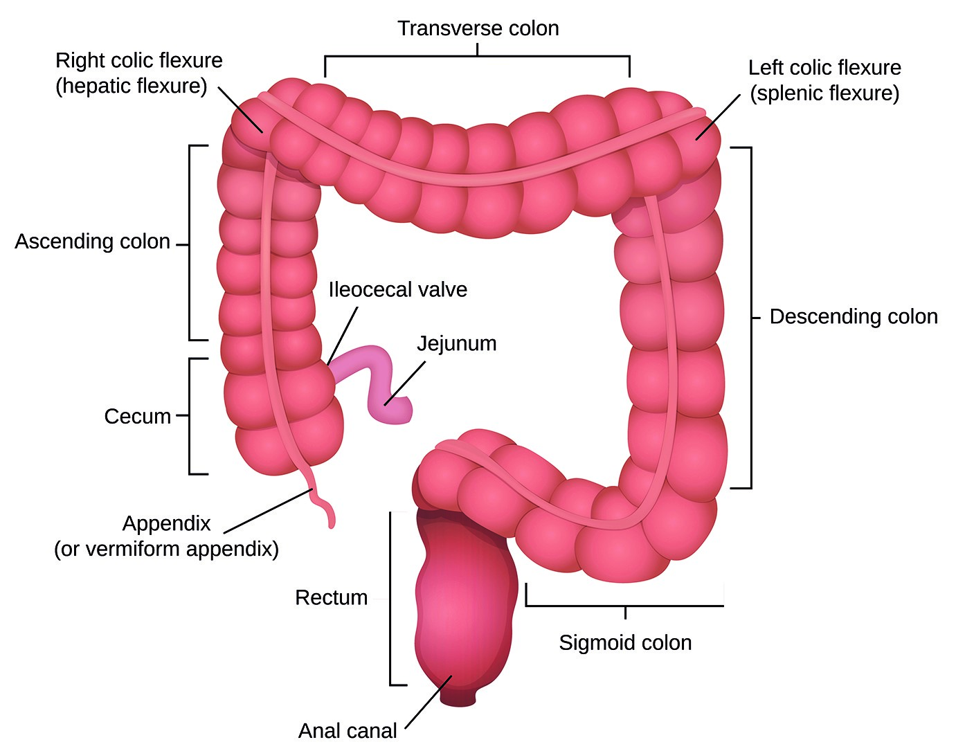
Figure 1. Anatomy of the large intestine.  

RECTUM, SPHINCTERS AND PELVIC FLOOR  


Rectum 

The primary function of the rectum is to collect and store faeces until being ready to defecate. Extrinsic autonomic nerves act upon the intrinsic nerves at submucosal plexuses to innervate the rectum. The combined effect of these nerves allows the rectal muscle the ability to relax and stretch and gradually accommodate increasing amounts of faecal content as it moves from the descending colon into the rectum (Norton and Chelvanayagam, 2004). This is known as rectal compliance. The rectum absorbs the remaining water, electrolytes and further solidifies waste products. The process of rectal emptying is usually initiated voluntarily. Anal rectal pressures are expressed in centimetres of water and resting pressure is low (between 5–20cm H2O) and does not increase significantly with increasing rectal content (Norton and Chelvanayagam, 2004). 
 

Internal and external anal sphincters  

The internal anal sphincter (IAS) is an involuntary circular smooth muscle approximately 0.3cm wide and ends 10mm above the anal verge. It is able to maintain tonic contraction for long periods of time and contributes to 85% of the resting anal tone, ranging from 60–110cm H2O (centimetres of water) in healthy individuals, and is under autonomic innervation. It is primarily responsible for closure of the anal canal at rest (Salvatore et al, 2017). The anorectal inhibitory reflex enables the internal sphincter to relax, allowing anal sensory receptors to sense rectal contents. This helps to differentiate solid or liquid stool from gas.  

The external anal sphincter (EAS; Figure 2) is a 0.6–1.0cm thick cylindrical striated muscle, which is under voluntary control (Salvatore et al, 2017). It surrounds the IAS and extends down to the anal verge. This muscle is fatigable and only contributes to 15% of the resting anal pressure tone. Its primary role is to preserve continence when stool or flatus is present in the rectum, or when intra-abdominal pressure rises, e.g. when coughing, sneezing, laughing. It relaxes to allow defaecation and is innervated by the inferior branch of the pudendal nerve (S3–S4) (Salvatore et al, 2017). 

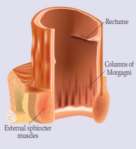
Figure 2. Rectum and internal and external sphincters.  

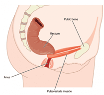
Figure 3. Puborectalis muscle contracted.  

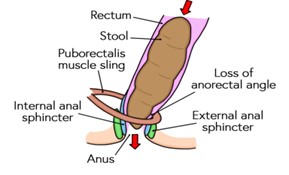
Figure 4. Puborectalis muscle at rest to allow passage of stool. 

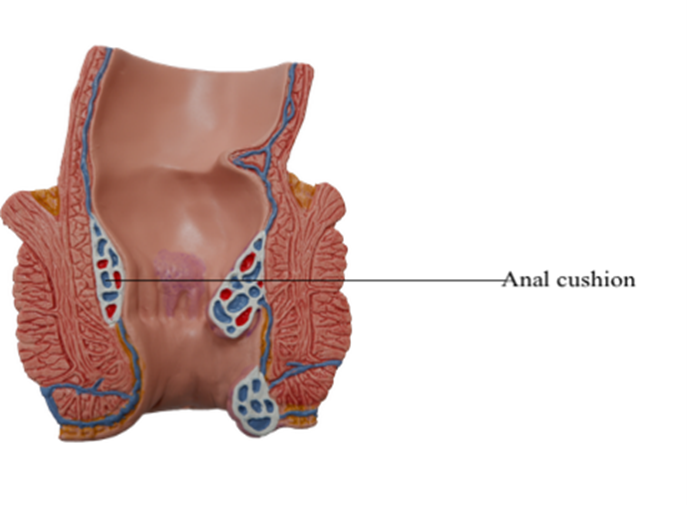
Figure 5. Anal cushions. 

Puborectalis muscle  


The puborectalis muscle (Figures 3 and 4) is part of the pelvic floor complex. It is a mixture of slow and fast twitch muscles and contains both types of muscle fibres. Its function is to close the upper anal canal and forms part of the anorectal angle. This angle is believed to be important in preserving continence and at rest forms an acute angle of 90 degrees, but during defaecation it becomes obtuse at an angle of between 110–130 degrees. This allows easier passage of the stool (Salvatore et al, 2017). The whole pelvic floor/anorectal angle mechanism works in conjunction with the anal sphincters during defaecation.
 

Anal cushions  


The submucosa of the anal lining contains blood vessels, connective tissue, smooth muscle and elastic tissue, which typically form three separate complexes of smooth muscle fibres and vascular channels called anal cushions (Figure 5). In the author’s clinical opinion, their contribution to continence is poorly studied and controversial, but they may assist with the closure mechanism by exerting pressures of up to 9mmHg, and thus possibly contributing 10–20% of resting anal pressure (Salvatore et al, 2017). 

STOOL PRODUCTION 


Normal stool output is about 150–200g per day. The upper colon defines consistency and volume of delivery of contents to the rectum. Bowel frequency in a healthy adult varies from three times daily to once every three days (Norton and Chelvanayagam, 2004; RCN, 2019). Faeces are made up of food residue, sloughed cells, unabsorbed gastrointestinal secretions, mucus and bacteria. Stool consistency can vary and has numerous influences, e.g. gender, diet and fluids.  

Defaecation or rectal emptying is usually initiated voluntarily. The rectum has a reservoir function to accommodate the contents until defaecation is appropriate. As rectal filling gradually proceeds ano rectal sampling takes place, with relaxation of the upper internal anal sphincter taking place approximately every 8–10 minutes. The contents are presented to the anal sensory mucosa in wave-like contractions, each lasting less than 10 seconds. This allows the sensory epithelium of the anal canal the opportunity to distinguish solids from liquid and gas. This is important in maintaining continence. Movement of the faeces into the rectum evokes the desire to defecate, known as the ‘call to stool’ (Norton and Chelvanayagam, 2004).  

When in a socially appropriate place, the person adopts a sitting/squatting position. This helps to straighten out the anorectal angle, allowing faeces to pass into the anal canal. Abdominal pressure is raised by contraction of the diaphragm and the abdominal muscles tensing to put pressure on pelvis. The puborectalis and external anal sphincter muscles relax, which allows for expulsion of the stool.  

Complete maintenance of continence is dependent on several major factors and is most likely with normal transit of formed stool (Table 1). 

In summary, the mechanism is as follows:   
  • Faeces enters rectum  
  • Reflex relaxation of sphincter  
  • Intra-abdominal pressure rises  
  • Anorectal angle straightens during sitting/squatting  
  • Gut contraction empties left colon into rectum  
  • Faeces squeezed out through anus. 
Table 1. Factors which maintain continence (adapted from Norton and Chelvanayagam, 2004; Salvatore et al, 2017).  
  • ● An effective barrier to outflow provided by an acute anorectal angle and anal sphincters 
  • ● Intact internal anal sphincters and anal cushions to ensure no passive leakage of stool  
  • ● Intact external anal sphincter to defer defaecation and reduce bowel urgency  
  • ● Intact rectal and anal sensation  
  • ● Compliant, distensible and evacuable reservoir (rectum)  
  • ● Intact central nervous system/functional reflexes for sampling stool 
  • ● Bulky and formed faeces   
  • ● Adequate cognitive (to recognise urge to defaecate) and physical mobility (to reach appropriate place). 

REFLEX ACTIONS 


There are a number of reflex actions with regards to the bowels that are also part of the anatomy and physiology. Healthcare professionals should be aware of these before any treatment option, namely: 
  • Gastro colic reflex — this is initiated when food and/or drink is ingested into the stomach and enhances muscular activity in the large bowel  
  • Anal reflex — when the skin around the anus is touched it contracts then relaxes. When undertaking certain procedures, e.g. suppositories/enemas use this reflex for easier insertion  
  • Closing reflex — this is the basis on which the anal sphincter snaps shut at the end of the evacuation process. This can be enhanced by squeezing the external sphincter muscle at the end of defaecation  
  • Recto anal inhibitory reflex — results when there is distension of the rectum where IAS relaxes and EAS contracts. This can be felt when the rectum fills and the ‘call to stool’ can be felt. 

CONCLUSION 


Faecal incontinence is prevalent and drastically affects quality of life. The anatomy and physiology of the bowels is complex and maintaining continence relies on all of these processes being intact. It is imperative that healthcare professionals not only understand the impact that this symptom has on individuals, but also comprehend how a normal bowel functions to determine what can go wrong. Part two of this series will explore bowel dysfunction and the causes of faecal incontinence, touching on how healthcare professionals can help. 
 
Ann Yates is Director of Continence Services, Cardiff and Vale UHB ann.yates@wales.nhs.uk 

References

Assmann SL, Keszthelyi D, Kleijnen J, et al (2022) Guideline for the diagnosis and treatment of Faecal Incontinence – A UEG/ESCP/ESNM/ESPCG collaboration. United European Gastroenterol J 10(3): 251-86  

Benezech A, Bouvier M, Vitton V (2016) Faecal incontinence: current knowledges and perspectives. World J Gastrointest Pathophysiol 7(1): 59-71 

Bliss DZ, Mimura T, Berghmans B, et al (2017) Assessment and conservative management of faecal incontinence and quality of life in adults. In: Abrams P, Cardozo L, Wagg A, Wein A, editors. Incontinence. 6th ed. International Continence Society, Bristol 

Camilleri-Brennan J (2020) Introductory chapter. Challenges in the diagnosis and treatment of faecal incontinence. In: Current Topics in Faecal Incontinence. IntechOpen. Available online: https://www.researchgate.net/publication/341185132_Introductory_Chapter_Challenges_in_the_Diagnosis_and_Treatment_of_Faecal_Incontinence  

Duelund-Jakobsen J, Worsoe J, Lundby L, Christensen P, Krough K (2016) Management of patients with faecal incontinence. Therap Adv Gastroenterol 9(1): 86-97 

Harari D (2009) Faecal incontinence in older people. Rev Clin Gerontol 19(2): 87-101  

International Continence Society (ICS) (2015) Fact Sheets. A Background to Urinary and Faecal Incontinence. Available online: https://www.ics.org/folder/82  

National Institute of Clinical Excellence (NICE) (2014) Faecal incontinence in adults. Quality Standard 54. Available online: www.nice.org.uk/guidance/qs54  

Norton C, Chelvanayagam S (2004) Bowel continence Nursing. Beaconsfield Publishers Ltd, Beaconsfield 

Norton C, Whitehead WE, Bliss DZ, Harari D, Lang J (2010) Management of faecal incontinence in adults. Neurourol Urodyn 29(1): 199-206  
 
Potter J, Peel P, Main S, et al (2007) National audit of continence care for older people: management of faecal incontinence. Age Ageing 36(3): 268-73  

Royal College of Nursing (2019) Bowel care. Management of lower bowel dysfunction, including digital rectal examination and digital removal of faeces. Available online: https://www.rcn.org.uk/professional-development/publications/pub-007522

Salvatore S, Delancey J, Igawa Y, et al (2017) Pathophysiology of Urinary incontinence, Faecal Incontinence and Pelvic organ prolapse. In: Incontinence 6th edition. (Eds) Abrams P, Cardozo L, Wagg A, Wein A. ICUD ICS, Tokyo 

Sbeit W, Khoury T, Mari A (2021) Diagnostic approach to faecal incontinence: what test and when to perform? World J Gastroenterol 27(15): 1553-62 

Young J (2022) Assessment and management of faecal incontinence. J Community Nurs 36(2): 51–8