

CHOOSING THE CORRECT CATHETER
There are many different types of indwelling urinary catheters available. The nurse should consider the use of latex, silicone, coated or composite materials, and pay attention to any patient history of sensitivity or allergy (Elvy and Colville, 2009).Latex catheters
Latex catheters are the most common type of catheter available. They are made from natural rubber and have been traditionally popular due to their flexibility. However, the high surface friction associated with latex can increase the risk of catheter encrustation, particularly around the catheter tip, which can increase pain and discomfort for the patient (Feneley et al, 2015; Yates, 2016). Sensitivity and allergy to latex is common and the initial assessment of each patient needs to consider the risks associated with the use of latex materials (Health and Safety Executive [HSE], 2011; NICE, 2017).Silicone catheters
 Silicone catheters have a wider internal lumen due to the composition, i.e. thinner walls than latex or coated catheters, and those made of 100% silicone are hypoallergenic for the majority of the population (Loveday et al, 2014). Silicone is slightly more rigid than latex and has a tendency to cause ‘cuffing’ or ‘ridging’ around the deflated balloon, which may in turn become attached to the urethral or suprapubic tract on removal, causing trauma (Geng et al, 2012; Feneley et al, 2015). In the author’s experience, this issue can be managed by completely deflating the catheter balloon and then inserting 1ml of sterile water to smooth out the edges of the balloon, which helps prevent the catheter from sticking to the urethral or abdominal tract tissues on removal. This, in turn, will reduce potential trauma.
Polytetrafluoroethylenecoated catheters
These are latex catheters coated in polytetrafluoroethylene [PTFE], which is smoother than latex and can be useful in reducing encrustation and discomfort for the wearer. There is still the risk of latex allergy and, therefore, PTFE-coated catheters must be avoided in patients with a known allergy or sensitivity.Hydrogel-coated catheters
Hydrogel-coated catheters can reduce friction and irritation, as they are soft,hydrophilic and biocompatible (Geng et al, 2012). They are typically a latex catheter with an integral hydrogel coating that offers a smooth catheter surface aimed at reducing friction and trauma on insertion.Silver-coated catheters
 Silver-coated catheters were introduced in the 1980s and are manufactured either as silicone, hydrogel or latex catheters with a thin layer silver alloy coating. Silver has long been recognised for its natural antimicrobial properties (Hayes, 2009), and these catheters were initially thought to significantly reduce the incidence of catheterassociated urinary tract infections (CAUTIs) (Pellowe, 2009; Stickler and Feneley, 2010; Pickard et al, 2012). However, it has since been acknowledged that the antimicrobial effect is short-lived and lasts no longer than 28 days (Tenke et al, 2008; Hayes, 2009; Beattie and Taylor, 2011; Geng et al, 2012; Loveday et al, 2014; Feneley et al, 2015). This means that the catheter has no added benefit in reducing infections after the 28 days and, therefore, would need changing to ensure optimum antimicrobial effect is continued. This will have cost implications, as these types of catheters are more expensive.
Nitrofurazone catheters
Nitrofurazone catheters are antibioticimpregnated catheters that may reduce asymptomatic bacteriuria when used as a short-term measure. However, the clinical evidence does not demonstrate a significant reduction in the incidence of symptomatic infection and, therefore, the use of this particular type of catheter is not widely recommended (Geng et al, 2012; Feneley et al, 2015).
BALLOON SIZE
 The balloon size of the catheter needs to be considered when choosing a product. Manufacturers produce Foley catheters with balloon sizes ranging from 5mls to 30mls. The larger balloons can increase the trauma and infection risk at the bladder neck, sphincter or suprapubic entry site, and leave a higher residual volume of urine within the bladder, thereby increasing the risk of infection from static urine (Garcia et al, 2007; Feneley et al, 2015). Larger balloons may also increase the incidence of urine bypassing, bladder spasms, pain and discomfort (Simpson, 2017). Larger balloon size catheters are commonest in urology when the Ch size is much larger, and the catheter is three-way rather than the standard two-way product to enable irrigation and effective drainage postoperatively. As the larger balloon size can cause trauma and increase the risk of infection, their use should be short term. A clinical indication for the choice of catheter should always be documented and the least risky option used.
Consideration should also be given to the loss of liquid volume that occurs over the catheter life of a sterile water-filled balloon, due to osmosis. While this is believed to be less of an issue when using glycerine solutions to inflate the balloon (Simpson, 2017), nurses should always follow the manufacturer’s advice when inflating a catheter balloon with water, and the balloon should never be overinflated.
CLEANSING SOLUTIONS
Indwelling urinary catheterisation is undertaken using an aseptic no-touch technique (ANTT) (Rowley et al, 2010; NICE, 2017). The use of sterile normal saline or sterile water to cleanse the meatal or abdominal skin is normally recommended before inserting the catheter (Healthcare Infection Control Practice Advisory Committee [HICPAC], 2009; Loveday et al, 2014). However, NICE (2017) has recommended that nurses use local policy and guidance for meatal cleansing before urethral catheterisation, without any specific reference to the type of cleansing solution.There have been some small studies reviewing the benefits of using an antimicrobial solution for meatal cleansing to reduce CAUTIs (Sandle, 2013; Levers, 2014). These have suggested that the use of such solutions may be beneficial, particularly in a community setting as the patient’s home environment can be more challenging from an infection control point of view, than a hospital setting. However, more work is required in this area before a definitive recommendation can be made on the advantages or disadvantages of introducing antimicrobial cleansing as a standard approach in urethral catheterisation. Until then, the best practice recommendation for managing catheter hygiene on a daily basis remains the use of soap and water in the individual’s normal personal hygiene routine (NICE, 2017).
LUBRICATION
Insertion of a urinary catheter, both suprapubic and urethral, requires the use of an appropriate lubricant so that trauma, discomfort and the risk of infection can be minimised (Loveday et al, 2014). Standard practice across the UK has been to use catheter lubricant gels that contain chlorhexidine (an antiseptic) or lidocaine (an anaesthetic), or a combination of both. However, in recent years there has been an increase in reports of adverse reactions, sensitivities and allergic reactions to chlorhexidine, possibly because of the inclusion of the active ingredient in many everyday household products (Marinho et al, 2013; Wilson, 2016).The clinical evidence suggesting that the use of chlorhexidine effectively reduces the incidence of CAUTIs is also limited and inconclusive (Wilson, 2016), with many countries switching to antiseptic-free gels (Williams, 2017). Several studies have outlined the benefits of anaesthetic gels in reducing pain on catheter insertion (Ramakrishnan and Mold, 2004; Kyle, 2009; Tzortzis et al, 2009).
During any assessment for catheterisation, it is the individual nurse’s responsibility to identify any contraindications, sensitivities or allergies that the patient may have to the active ingredients used in lubricating gels. Similarly, the nurse should use the assessment to choose an appropriate catheter for the patient that will ensure that the experience of catheterisation is as safe, effective and comfortable as possible.
CATHETER-ASSOCIATED URINARY TRACT INFECTIONS
CAUTIs account for a large proportion of healthcare-acquired infections (Pellowe, 2009) and the cost of treating a single CAUTI is estimated at almost £2,000 (Loveday et al, 2014), placing an enormous burden on the healthcare economy. Establishing the effect of a CAUTI on a patient’s quality of life is difficult to determine, with the risk of serious infection rising the longer the catheter is in place (Chang et al, 2011; Loveday et al, 2014). Forty-five per cent Escherichia coli bacteraemia are attributed to the urinary tract and use of catheters (Abernathy, 2017). It is now a Public Health England policy to reduce all healthcare-associated Gram-negative bloodstream infections by 50% by 2021, and all trusts have been challenged with ensuring a robust action plan is in place to achieve this by closer monitoring, robust early detection and appropriate treatment of CAUTIs (NHS Improvement, 2017).ENCRUSTATION
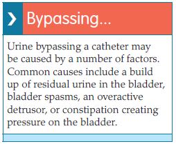
Catheter blockage and bypassing are common issues encountered with the use of indwelling urinary catheters and are usually caused by infection and encrustation. Encrustation is commonly caused by a build-up of Proteus mirabilis, a urease-producing bacteria, which causes biofilm formation on the catheter surface leading to blockage of the lumen and drainage eyelets (Stickler et al, 2003; Feneley et al, 2015). Traditionally, catheter maintenance solutions have routinely been used to dissolve the encrustation or remove debris. However, this is a high-risk strategy for a number of reasons (Turner and Dickens, 2011; Davey, 2015; Feneley et al, 2015; Gibney, 2016):- To flush the catheter with maintenance solutions requires breaking the closed drainage system, thus increasing the risk of infection
- The acidic content of catheter maintenance solutions can damage the urothelial lining of the bladder and cause an inflammatory response
- The increased pressure under which they can be administered, can also contribute to significant damage and increased infection risk. Thus, pre-filled catheter maintainence solutions should be gravity fed/administered. Any squeezing of the container/bag will create pressure within the catheter and bladder which can cause trauma and tissue damage. Catheter maintenance solutions should be used with caution, and only after a thorough assessment of risk and the formulation of a clear clinical rationale, which must be documented in the patient’s records.
Antibiotics and antiseptics are ineffective in reducing catheter encrustation; however, antimicrobials used in the ballooninflation solution may be effective in reducing harmful bacteria including P. mirabilis (Pannek and Vesweber, 2011; Sperling et al, 2014; NICE, 2017).
References
Abernathy J, Guy R, Sheridan EA, et al (2017) Epidemiology of Escherichia coli bacteraemia in England, Results of an enhanced sentinel surveillance programme. J Hosp Infect 95(4): 365–75
Beattie M, Taylor J (2011) Silver alloy versus uncoated urinary catheters: a systematic review of the literature. J Clin Nurs 20: 2098–2108
Chang R, Todd GM, Chenoweth CE (2011) Epidemiology of hospital acquired urinary tract related bloodstream infection at a university hospital. Infect Contr Hosp Epidemiol 32(11): 1127–9
Davey G (2015) Troubleshooting indwelling catheter problems in the community. J Community Nurs 29(4): 67–74
Elvy J, Colville A (2009) Catheter associated urinary tract infection: what is it, what causes it, and how can we prevent it? J Infect Prevent 10(2): 36–41
Feneley R, Hopley I, Wells P (2015) Urinary catheters history: current status, adverse events and research agenda. J Med Eng Technol 39(8): 459–70
Garcia MM, Gulati S, Liepmann D, et al (2007) Traditional Foley drainage systems — do they drain the bladder? J Urol 177: 203–7
Geng V, Cobussen-Boekhorst H, Farrell J (2012) Catheterisation: Indwelling in Adults. European Association of Urology Nurses (EAUN), Holland
Gibney LE (2016) Blocked urinary catheters: can they be better managed? Br J Nurs 25(15): 828-33
Hanchett M (2002) Techniques for stabilising urinary catheters: tape may be the oldest method but it’s not the only one. Am J Nurs 102(3): 44–48
Hayes W (2009) Looking past the silver lining. Materials Management in Healthcare January: 20–23. Available online: matmanmag.com
Healthcare Infection Control Practices Advisory Committee (2009) Guideline for Prevention of Catheter-associated Urinary Tract Infection. Available online: www.cdc.gov/infectioncontrol/pdf/ guidelines/cauti-guidelines.pdf (accessed 1 February, 2018)
Health Protection Scotland (2012) Scottish National Point Prevalence Survey of Healthcare Associated Infection and Antimicrobial Prescribing 2011. Available online: http://bit.ly/1SJCIcK
Health and Safety Executive (2011) Natural rubber latex sensitisation in health and social care. Available online: www.hse.gov. uk/foi/internalops/sims/pub_serv/071106. htm (accessed 30 January, 2018)
Holroyd S (2016) Innovation in catheter securement devices: minimising risk of infection, trauma and pain. Br J Community Nurs 21(5): 256–9
Kyle G (2009) Lubricating gels in urethral catheterisation — the evidence. Continence UK 3(1): 35–7
Leaver R (2017) Understanding long-term catheterisation for effective bladder drainage. J Community Nurs 31(5): 43–8
Levers H (2014) Switching to an antimicrobial solution for skin cleansing before urinary catheterisation. Br J Community Nurs 19(2): 66–71
Loveday HP, Wilson JA, Pratt RJ (2014) EPIC 3: national evidence-based guidelines for preventing healthcare associated infections in NHS hospitals in England. J Hosp Infect 86(Suppl 1): S1–S70
Marinho S, Oliver P, Hughes D (2013) Chlorhexidine Allergy Policy: Screening and Management of Patients with Chlorhexidine Allergy. University Hospital of South Manchester NHS Foundation Trust, Manchester
National Institute for Health and Care Excellence (2014) Long Term Urinary Catheters: Prevention and Control of Healthcare-associated Infections in Primary and Community Care. NICE, London
National Institute for Health and Care Excellence (2017) Healthcare-associated Infections: Prevention and Control in Primary and Community Care. Clinical guideline 139. NICE, London
National Patient Safety Agency (2009) Rapid Response Report: minimising risks of suprapubic catheter insertion (adults only). Available online: www.nrls.npsa.nhs.uk/ alerts/?entryid45=61917 (accessed 2 February, 2018)
NHS Improvement (2017) Preventing Healthcare-associated Gram-negative Bloodstream Infections: An Improvement Resource. Public Health England, London
Pannek J, Vesweber A (2011) Clinical utility of an antimicrobial blocking solution in patients with an indwelling catheter. Aktuelle Urol 42: 51–4
Pellowe C (2009) EPIC 2 infection prevention guidelines — best practice. Continence UK 3(3): 28–33
Pickard R, Lam T, MacLennan GL (2012) Antimicrobial catheters for reduction of symptomatic urinary tract infection in adults requiring short-term catheterisation in hospital: a multicentre randomised controlled trial. Lancet 380(9857): 1927–35
Ramakrishnan K, Mold J (2004) Urinary catheters: a review. Int J Fam Pract. Available online: http://ispub.com/ IJFP/3/2/4596
Rowley S, Clare S, Macqueen S, Molyneux R (2010) ANTT v2: an updated practice framework for aseptic technique. Br J Nurs 19(5 Suppl): S5–11
Royal College of Nursing (2012) Catheter Care: RCN Guidance for Nurses. RCN, London Royal College of Physicians (2005) Report of the National Audit of Continence Care in Older People (65 years and above) in England, Wales and Northern Ireland. Royal College of Physicians, London
Sandle T (2013) Using an antimicrobial skin cleanser before catheterisation. J Community Nurs 27(5): 30–4
Simpson P (2017) Long-term urethral catheterisation: guidelines for community nurses. Br J Nurs 26(9 Suppl): S22–S26
Sperling H, Eisenhardt A, Mumperow E (2014) Review of the use of triclosan in permanent catheters. Der Urologe 53: 1512–17
Spinks J (2013) Urinary incontinence and the importance of catheter fixation. J Community Nurs 27(5 Suppl): S24–S29
Stickler D, Young R, Jones G, Sabbuba N, Morris N (2003) Why are foley catheters so vulnerable to encrustation and blockage by crystalline bacterial biofilm? Urol Res 31: 306–11
Stickler DJ, Feneley RCL (2010) The encrustation and blockage of longterm indwelling bladder catheters: a way forward in prevention and control. Spinal Cord 48(11): 784–90
Tenke P, Kovacs B, Bjerklund Johansen TE (2008) European and Asian guidelines on management and prevention of catheter-associated urinary tract infections. Int J Antimicrob Agents 31(1 Suppl): S68–78
Turner B, Dickens N (2011) Long-term urethral catheterisation. Nurs Standard 25(24): 49–55
Tzortzis V, Gravas S, Melekos MM, de la Rosette JJ (2009) Intraurethral lubricants: a critical literature review and recommendations. J Endourol 23(5): 821–6
Williams C (2017) Making a choice of catheterisation gel and the role of chlorhexidine. Br J Community Nurs 22(7): 346–51
Wilson M (2016) Urinary catheterisation in the community: exploring challenges and solutions. Br J Community Nurs 21(10): 443
Woodward S (2014) Securing urethral catheters can help to reduce their complications. Br J Neuro Nurs 10(4): 162–5
Wound, Ostomy and Continence Nurses Society (2012) Indwelling Urinary Catheter Securement: Best Practice for Clinicians. WOCN, USA
Yarde D (2015) Managing indwelling urinary catheters in adults. Nurs Times 111(22): 12–13
Yates A (2016) Indwelling urinary catheterisation: what is best practice? Br J Nurs 25(9 Suppl): S4–12